3CCH | pdb_00003cch

H-2Db complex with murine gp100


Experimental Data Snapshot

  • Method: X-RAY DIFFRACTION
  • Resolution: 2.60 Å
  • R-Value Free: 
    0.279 (Depositor), 0.274 (DCC) 
  • R-Value Work: 
    0.241 (Depositor), 0.239 (DCC) 
  • R-Value Observed: 
    0.243 (Depositor) 

wwPDB Validation 3D Report Full Report

Validation slider image for 3CCH

This is version 1.2 of the entry. See complete history

Literature

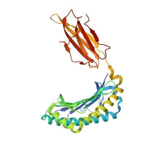
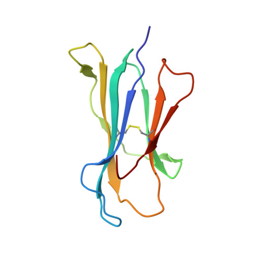
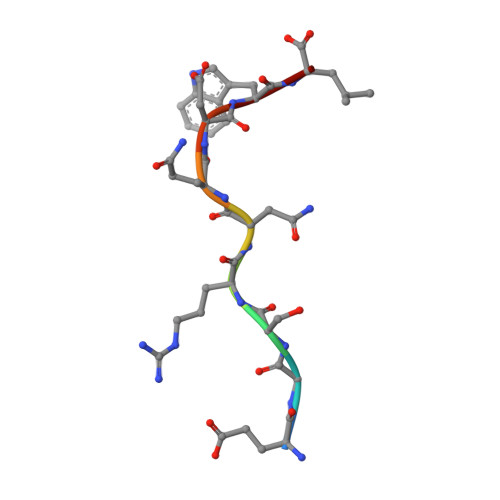
Design of agonistic altered peptides for the robust induction of CTL directed towards H-2Db in complex with the melanoma-associated epitope gp100.

van Stipdonk, M.J.Badia-Martinez, D.Sluijter, M.Offringa, R.van Hall, T.Achour, A.

(2009) Cancer Res 69: 7784-7792

  • DOI: https://doi.org/10.1158/0008-5472.CAN-09-1724
  • Primary Citation Related Structures: 
    3CC5, 3CCH, 3CH1

  • PubMed Abstract: 

    Immunogenicity of tumor-associated antigens (TAA) is often weak because many TAA are autoantigens for which the T-cell repertoire is sculpted by tolerance mechanisms. Substitutions at main anchor positions to increase the complementarity between the peptide and the MHC class I (MHC-I) binding cleft constitute a common procedure to improve binding capacity and immunogenicity of TAA. However, such alterations are tailored for each MHC-I allele and may recruit different CTL specificities through conformational changes in the targeted peptides. Comparative analysis of substituted melanoma-differentiation antigen gp100 in complex with H-2D(b) revealed that combined introduction of glycine and proline residues at the nonanchor positions 2 and 3, respectively, resulted in an agonistic altered peptide with dramatically enhanced binding affinity, stability, and immunogenicity of this TAA. Peptide vaccination using the p2Gp3P-altered peptide version of gp100 induced high frequencies of melanoma-specific CTL in the endogenous CD8+ repertoire. Crystal structure analysis of MHC/peptide complexes revealed that the conformation of the modified p2Gp3P-peptide was similar to the wild-type peptide, and indicated that this mimotope was stabilized through interactions between peptide residue p3P and the tyrosine residue Y159 that is conserved among most known MHC-I molecules throughout mammalian species. Our results may provide an alternative approach to enhance MHC stabilization capacity and immunogenicity of low-affinity peptides for induction of robust tumor-specific CTL.


  • Organizational Affiliation
    • Departments of Immunohematology and Blood transfusion and Clinical Oncology, Leiden University Medical Center, Leiden, the Netherlands.

Macromolecule Content 

  • Total Structure Weight: 181.97 kDa 
  • Atom Count: 12,836 
  • Modeled Residue Count: 1,528 
  • Deposited Residue Count: 1,536 
  • Unique protein chains: 3

Macromolecules

Find similar proteins by:|  3D Structure
Entity ID: 1
MoleculeChains  Sequence LengthOrganismDetailsImage
H-2 class I histocompatibility antigen, D-B alpha chain
A, D, G, J
276Mus musculusMutation(s): 0 
Gene Names: H2-D1
UniProt
Find proteins for P01899 (Mus musculus)
Explore P01899 
Go to UniProtKB:  P01899
Entity Groups
Sequence Clusters30% Identity50% Identity70% Identity90% Identity95% Identity100% Identity
UniProt GroupP01899
Sequence Annotations
Expand
Reference Sequence
Find similar proteins by:|  3D Structure
Entity ID: 2
MoleculeChains  Sequence LengthOrganismDetailsImage
Beta-2-microglobulin
B, E, H, K
99Mus musculusMutation(s): 0 
Gene Names: B2M
UniProt & NIH Common Fund Data Resources
Find proteins for P01887 (Mus musculus)
Explore P01887 
Go to UniProtKB:  P01887
IMPC:  MGI:88127
Entity Groups
Sequence Clusters30% Identity50% Identity70% Identity90% Identity95% Identity100% Identity
UniProt GroupP01887
Sequence Annotations
Expand
Reference Sequence
Find similar proteins by:  Sequence   |   3D Structure  
Entity ID: 3
MoleculeChains  Sequence LengthOrganismDetailsImage
nonameric peptide murine gp100
C, F, I, L
9N/AMutation(s): 0 

Small Molecules

Ligands 2 Unique
IDChains Name / Formula / InChI Key2D Diagram3D Interactions
SO4

Query on SO4


Download:Ideal Coordinates CCD File
BA [auth H]
CA [auth H]
DA [auth H]
EA [auth H]
FA [auth J]
BA [auth H],
CA [auth H],
DA [auth H],
EA [auth H],
FA [auth J],
GA [auth J],
IA [auth K],
JA [auth K],
KA [auth K],
N [auth B],
O [auth B],
P [auth B],
Q [auth B],
R [auth D],
S [auth D],
U [auth E],
V [auth E],
W [auth E],
X [auth E],
Y [auth E],
Z [auth G]
SULFATE ION
O4 S
QAOWNCQODCNURD-UHFFFAOYSA-L
GOL

Query on GOL


Download:Ideal Coordinates CCD File
AA [auth G],
HA [auth J],
M [auth A],
T [auth D]
GLYCEROL
C3 H8 O3
PEDCQBHIVMGVHV-UHFFFAOYSA-N

Experimental Data & Validation

Experimental Data

  • Method: X-RAY DIFFRACTION
  • Resolution: 2.60 Å
  • R-Value Free:  0.279 (Depositor), 0.274 (DCC) 
  • R-Value Work:  0.241 (Depositor), 0.239 (DCC) 
  • R-Value Observed: 0.243 (Depositor) 
Space Group: P 1 21 1
Unit Cell:
Length ( Å )Angle ( ˚ )
a = 85.5α = 90
b = 176.6β = 120
c = 85.5γ = 90
Software Package:
Software NamePurpose
REFMACrefinement
ADSCdata collection
XDSdata reduction
XSCALEdata scaling
PHASERphasing

Structure Validation

View Full Validation Report


Entry History 

Deposition Data

Revision History  (Full details and data files)

  • Version 1.0: 2009-03-10
    Type: Initial release
  • Version 1.1: 2011-07-13
    Changes: Non-polymer description, Version format compliance
  • Version 1.2: 2024-10-16
    Changes: Data collection, Database references, Derived calculations, Refinement description, Structure summary