3CCN | pdb_00003ccn

X-ray structure of c-Met with triazolopyridazine inhibitor.


Experimental Data Snapshot

  • Method: X-RAY DIFFRACTION
  • Resolution: 1.90 Å
  • R-Value Free: 
    0.275 (Depositor), 0.265 (DCC) 
  • R-Value Work: 
    0.236 (Depositor), 0.230 (DCC) 
  • R-Value Observed: 
    0.238 (Depositor) 

Starting Model: experimental
View more details

wwPDB Validation 3D Report Full Report

Validation slider image for 3CCN

Ligand Structure Quality Assessment 


This is version 1.2 of the entry. See complete history

Literature

Discovery and Optimization of Triazolopyridazines as Potent and Selective Inhibitors of the c-Met Kinase.

Albrecht, B.K.Harmange, J.C.Bauer, D.Berry, L.Bode, C.Boezio, A.A.Chen, A.Choquette, D.Dussault, I.Fridrich, C.Hirai, S.Hoffman, D.Larrow, J.F.Kaplan-Lefko, P.Lin, J.Lohman, J.Long, A.M.Moriguchi, J.O'Connor, A.Potashman, M.H.Reese, M.Rex, K.Siegmund, A.Shah, K.Shimanovich, R.Springer, S.K.Teffera, Y.Yang, Y.Zhang, Y.Bellon, S.F.

(2008) J Med Chem 51: 2879-2882

  • DOI: https://doi.org/10.1021/jm800043g
  • Primary Citation Related Structures: 
    3CCN, 3CD8

  • PubMed Abstract: 

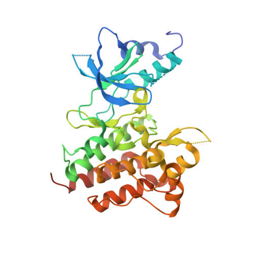
    Tumorigenesis is a multistep process in which oncogenes play a key role in tumor formation, growth, and maintenance. MET was discovered as an oncogene that is activated by its ligand, hepatocyte growth factor. Deregulated signaling in the c-Met pathway has been observed in multiple tumor types. Herein we report the discovery of potent and selective triazolopyridazine small molecules that inhibit c-Met activity.


  • Organizational Affiliation
    • Amgen Inc., Cambridge, MA 02139, USA. brian.albrecht@amgen.com

Macromolecule Content 

  • Total Structure Weight: 35.65 kDa 
  • Atom Count: 2,348 
  • Modeled Residue Count: 275 
  • Deposited Residue Count: 310 
  • Unique protein chains: 1

Macromolecules

Find similar proteins by:|  3D Structure
Entity ID: 1
MoleculeChains  Sequence LengthOrganismDetailsImage
Hepatocyte growth factor receptor310Homo sapiensMutation(s): 1 
Gene Names: MET
EC: 2.7.10.1
UniProt & NIH Common Fund Data Resources
Find proteins for P08581 (Homo sapiens)
Explore P08581 
Go to UniProtKB:  P08581
PHAROS:  P08581
GTEx:  ENSG00000105976 
Entity Groups
Sequence Clusters30% Identity50% Identity70% Identity90% Identity95% Identity100% Identity
UniProt GroupP08581
Sequence Annotations
Expand
Reference Sequence

Small Molecules

Ligands 1 Unique
IDChains Name / Formula / InChI Key2D Diagram3D Interactions
LKG

Query on LKG


Download:Ideal Coordinates CCD File
B [auth A]4-[(6-phenyl[1,2,4]triazolo[4,3-b]pyridazin-3-yl)methyl]phenol
C18 H14 N4 O
ZGJYGQLGSXWEMY-UHFFFAOYSA-N

Experimental Data & Validation

Experimental Data

  • Method: X-RAY DIFFRACTION
  • Resolution: 1.90 Å
  • R-Value Free:  0.275 (Depositor), 0.265 (DCC) 
  • R-Value Work:  0.236 (Depositor), 0.230 (DCC) 
  • R-Value Observed: 0.238 (Depositor) 
Space Group: P 21 21 21
Unit Cell:
Length ( Å )Angle ( ˚ )
a = 42.058α = 90
b = 43.44β = 90
c = 157.827γ = 90
Software Package:
Software NamePurpose
REFMACrefinement
CrystalCleardata collection
DENZOdata reduction
SCALEPACKdata scaling
AMoREphasing

Structure Validation

View Full Validation Report


Ligand Structure Quality Assessment 


Entry History 

Deposition Data

Revision History  (Full details and data files)

  • Version 1.0: 2008-04-29
    Type: Initial release
  • Version 1.1: 2011-07-13
    Changes: Version format compliance
  • Version 1.2: 2023-08-30
    Changes: Data collection, Database references, Derived calculations, Refinement description