3CLD

Ligand binding domain of the glucocorticoid receptor complexed with fluticazone furoate


Experimental Data Snapshot

  • Method: X-RAY DIFFRACTION
  • Resolution: 2.84 Å
  • R-Value Free: 0.267 
  • R-Value Work: 0.207 
  • R-Value Observed: 0.210 

wwPDB Validation   3D Report Full Report


Ligand Structure Quality Assessment 


This is version 1.3 of the entry. See complete history


Literature

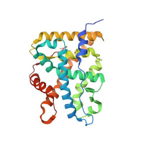
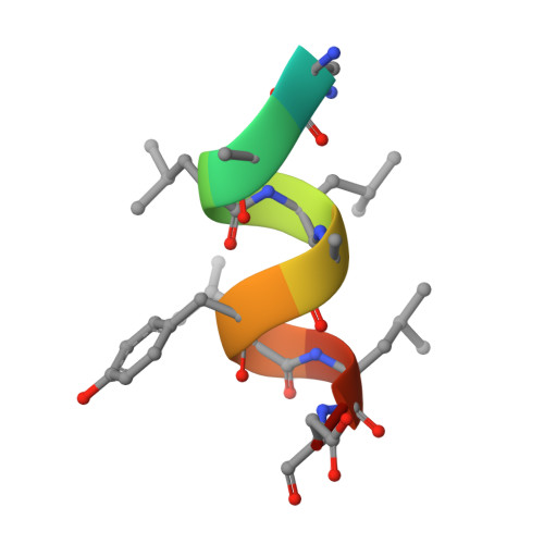
X-ray crystal structure of the novel enhanced-affinity glucocorticoid agonist fluticasone furoate in the glucocorticoid receptor-ligand binding domain.

Biggadike, K.Bledsoe, R.K.Hassell, A.M.Kirk, B.E.McLay, I.M.Shewchuk, L.M.Stewart, E.L.

(2008) J Med Chem 51: 3349-3352

  • DOI: https://doi.org/10.1021/jm800279t
  • Primary Citation of Related Structures:  
    3CLD

  • PubMed Abstract: 

    An X-ray crystal structure is reported for the novel enhanced-affinity glucocorticoid agonist fluticasone furoate (FF) in the ligand binding domain of the glucocorticoid receptor. Comparison of this structure with those of dexamethasone and fluticasone propionate shows the 17 alpha furoate ester to occupy more fully the lipophilic 17 alpha pocket on the receptor, which may account for the enhanced glucocorticoid receptor binding of FF.


  • Organizational Affiliation

    kb0903@gsk.com


Macromolecules
Find similar proteins by:  (by identity cutoff)  |  3D Structure
Entity ID: 1
MoleculeChains Sequence LengthOrganismDetailsImage
Glucocorticoid receptorA,
C [auth B]
259Homo sapiensMutation(s): 2 
Gene Names: NR3C1GRL
UniProt & NIH Common Fund Data Resources
Find proteins for P04150 (Homo sapiens)
Explore P04150 
Go to UniProtKB:  P04150
PHAROS:  P04150
GTEx:  ENSG00000113580 
Entity Groups  
Sequence Clusters30% Identity50% Identity70% Identity90% Identity95% Identity100% Identity
UniProt GroupP04150
Sequence Annotations
Expand
  • Reference Sequence

Find similar proteins by:  Sequence   |   3D Structure  

Entity ID: 2
MoleculeChains Sequence LengthOrganismDetailsImage
Tif2 coactivator motifB [auth H],
D [auth C]
12N/AMutation(s): 0 
UniProt & NIH Common Fund Data Resources
Find proteins for Q15596 (Homo sapiens)
Explore Q15596 
Go to UniProtKB:  Q15596
PHAROS:  Q15596
GTEx:  ENSG00000140396 
Entity Groups  
Sequence Clusters30% Identity50% Identity70% Identity90% Identity95% Identity100% Identity
UniProt GroupQ15596
Sequence Annotations
Expand
  • Reference Sequence
Small Molecules
Ligands 1 Unique
IDChains Name / Formula / InChI Key2D Diagram3D Interactions
GW6
Query on GW6

Download Ideal Coordinates CCD File 
E [auth A],
F [auth B]
(6alpha,11alpha,14beta,16alpha,17alpha)-6,9-difluoro-17-{[(fluoromethyl)sulfanyl]carbonyl}-11-hydroxy-16-methyl-3-oxoan drosta-1,4-dien-17-yl furan-2-carboxylate
C27 H29 F3 O6 S
XTULMSXFIHGYFS-VLSRWLAYSA-N
Binding Affinity Annotations 
IDSourceBinding Affinity
GW6 BindingDB:  3CLD Ki: 0.51 (nM) from 1 assay(s)
IC50: min: 0.04, max: 0.8 (nM) from 2 assay(s)
EC50: min: 0.4, max: 0.49 (nM) from 2 assay(s)
Experimental Data & Validation

Experimental Data

  • Method: X-RAY DIFFRACTION
  • Resolution: 2.84 Å
  • R-Value Free: 0.267 
  • R-Value Work: 0.207 
  • R-Value Observed: 0.210 
  • Space Group: P 61
Unit Cell:
Length ( Å )Angle ( ˚ )
a = 127.34α = 90
b = 127.34β = 90
c = 77.772γ = 120
Software Package:
Software NamePurpose
REFMACrefinement
HKL-2000data collection
HKL-2000data reduction
HKL-2000data scaling
CNSphasing

Structure Validation

View Full Validation Report


Ligand Structure Quality Assessment 


Entry History 

Deposition Data

Revision History  (Full details and data files)

  • Version 1.0: 2008-06-24
    Type: Initial release
  • Version 1.1: 2011-07-13
    Changes: Advisory, Version format compliance
  • Version 1.2: 2021-10-20
    Changes: Database references, Derived calculations, Structure summary
  • Version 1.3: 2023-08-30
    Changes: Data collection, Refinement description