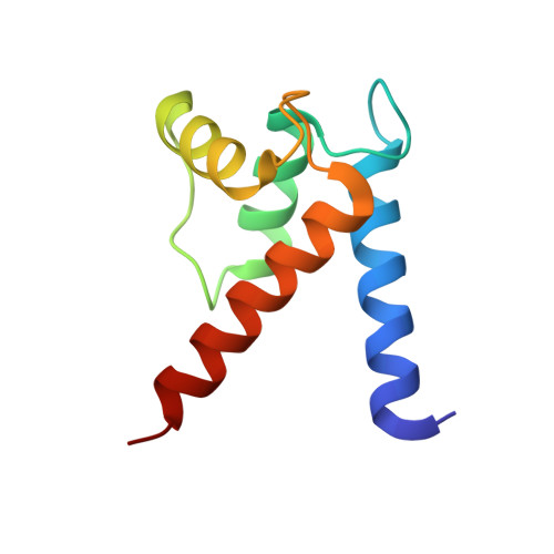
3CR5

X-ray structure of bovine Pnt-Zn(2+),Ca(2+)-S100B


Experimental Data Snapshot

  • Method: X-RAY DIFFRACTION
  • Resolution: 1.85 Å
  • R-Value Free: 0.243 
  • R-Value Work: 0.192 
  • R-Value Observed: 0.194 

wwPDB Validation   3D Report Full Report


Ligand Structure Quality Assessment 


This is version 1.3 of the entry. See complete history


Literature

Divalent metal ion complexes of S100B in the absence and presence of pentamidine.

Charpentier, T.H.Wilder, P.T.Liriano, M.A.Varney, K.M.Pozharski, E.MacKerell, A.D.Coop, A.Toth, E.A.Weber, D.J.

(2008) J Mol Biol 382: 56-73

  • DOI: https://doi.org/10.1016/j.jmb.2008.06.047
  • Primary Citation of Related Structures:  
    3CR2, 3CR4, 3CR5

  • PubMed Abstract: 

    As part of an effort to inhibit S100B, structures of pentamidine (Pnt) bound to Ca(2+)-loaded and Zn(2+),Ca(2+)-loaded S100B were determined by X-ray crystallography at 2.15 A (R(free)=0.266) and 1.85 A (R(free)=0.243) resolution, respectively. These data were compared to X-ray structures solved in the absence of Pnt, including Ca(2+)-loaded S100B and Zn(2+),Ca(2+)-loaded S100B determined here (1.88 A; R(free)=0.267). In the presence and absence of Zn(2+), electron density corresponding to two Pnt molecules per S100B subunit was mapped for both drug-bound structures. One Pnt binding site (site 1) was adjacent to a p53 peptide binding site on S100B (+/-Zn(2+)), and the second Pnt molecule was mapped to the dimer interface (site 2; +/-Zn(2+)) and in a pocket near residues that define the Zn(2+) binding site on S100B. In addition, a conformational change in S100B was observed upon the addition of Zn(2+) to Ca(2+)-S100B, which changed the conformation and orientation of Pnt bound to sites 1 and 2 of Pnt-Zn(2+),Ca(2+)-S100B when compared to Pnt-Ca(2+)-S100B. That Pnt can adapt to this Zn(2+)-dependent conformational change was unexpected and provides a new mode for S100B inhibition by this drug. These data will be useful for developing novel inhibitors of both Ca(2+)- and Ca(2+),Zn(2+)-bound S100B.


  • Organizational Affiliation

    Department of Biochemistry and Molecular Biology, The University of Maryland School of Medicine, 108 North Greene Street, Baltimore, MD 21201, USA.


Macromolecules
Find similar proteins by:  (by identity cutoff)  |  3D Structure
Entity ID: 1
MoleculeChains Sequence LengthOrganismDetailsImage
Protein S100-BA [auth X]92Bos taurusMutation(s): 0 
Gene Names: S100B
UniProt
Find proteins for P02638 (Bos taurus)
Explore P02638 
Go to UniProtKB:  P02638
Entity Groups  
Sequence Clusters30% Identity50% Identity70% Identity90% Identity95% Identity100% Identity
UniProt GroupP02638
Sequence Annotations
Expand
  • Reference Sequence
Small Molecules
Binding Affinity Annotations 
IDSourceBinding Affinity
PNT BindingDB:  3CR5 Kd: min: 1000, max: 5.00e+4 (nM) from 3 assay(s)
IC50: 1.00e+5 (nM) from 1 assay(s)
PDBBind:  3CR5 Kd: 6.40e+4 (nM) from 1 assay(s)
Experimental Data & Validation

Experimental Data

  • Method: X-RAY DIFFRACTION
  • Resolution: 1.85 Å
  • R-Value Free: 0.243 
  • R-Value Work: 0.192 
  • R-Value Observed: 0.194 
  • Space Group: P 41 21 2
Unit Cell:
Length ( Å )Angle ( ˚ )
a = 62.883α = 90
b = 62.883β = 90
c = 50.084γ = 90
Software Package:
Software NamePurpose
DENZOdata reduction
SCALEPACKdata scaling
MOLREPphasing
REFMACrefinement
PDB_EXTRACTdata extraction
Blu-Icedata collection

Structure Validation

View Full Validation Report


Ligand Structure Quality Assessment 


Entry History 

Deposition Data

Revision History  (Full details and data files)

  • Version 1.0: 2008-06-24
    Type: Initial release
  • Version 1.1: 2011-07-13
    Changes: Advisory, Version format compliance
  • Version 1.2: 2017-10-25
    Changes: Refinement description
  • Version 1.3: 2024-02-21
    Changes: Data collection, Database references, Derived calculations