3CW3 | pdb_00003cw3

Crystal structure of AIM1g1


Experimental Data Snapshot

  • Method: X-RAY DIFFRACTION
  • Resolution: 1.88 Å
  • R-Value Free: 
    0.232 (Depositor), 0.181 (DCC) 
  • R-Value Work: 
    0.181 (Depositor), 0.178 (DCC) 

wwPDB Validation 3D Report Full Report

Validation slider image for 3CW3

This is version 1.3 of the entry. See complete history

Literature

Exploring the limits of sequence and structure in a variant betagamma-crystallin domain of the protein absent in melanoma-1 (AIM1).

Aravind, P.Wistow, G.Sharma, Y.Sankaranarayanan, R.

(2008) J Mol Biology 381: 509-518

  • DOI: https://doi.org/10.1016/j.jmb.2008.06.019
  • Primary Citation Related Structures: 
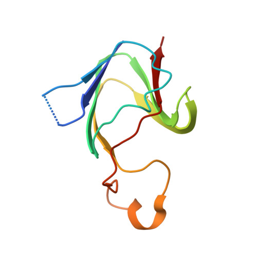
    3CW3

  • PubMed Abstract: 

    Betagamma-crystallins belong to a superfamily of proteins in prokaryotes and eukaryotes that are based on duplications of a characteristic, highly conserved Greek key motif. Most members of the superfamily in vertebrates are structural proteins of the eye lens that contain four motifs arranged as two structural domains. Absent in melanoma 1 (AIM1), an unusual member of the superfamily whose expression is associated with suppression of malignancy in melanoma, contains 12 betagamma-crystallin motifs in six domains. Some of these motifs diverge considerably from the canonical motif sequence. AIM1g1, the first betagamma-crystallin domain of AIM1, is the most variant of betagamma-crystallin domains currently known. In order to understand the limits of sequence variation on the structure, we report the crystal structure of AIM1g1 at 1.9 A resolution. Despite having changes in key residues, the domain retains the overall betagamma-crystallin fold. The domain also contains an unusual extended surface loop that significantly alters the shape of the domain and its charge profile. This structure illustrates the resilience of the betagamma fold to considerable sequence changes and its remarkable ability to adapt for novel functions.


  • Organizational Affiliation
    • Centre for Cellular and Molecular Biology, Uppal Road, Hyderabad-500007, India.

Macromolecule Content 

  • Total Structure Weight: 10.91 kDa 
  • Atom Count: 856 
  • Modeled Residue Count: 93 
  • Deposited Residue Count: 96 
  • Unique protein chains: 1

Macromolecules

Find similar proteins by:|  3D Structure
Entity ID: 1
MoleculeChains  Sequence LengthOrganismDetailsImage
Absent in melanoma 1 protein96Homo sapiensMutation(s): 0 
Gene Names: AIM1
UniProt & NIH Common Fund Data Resources
Find proteins for Q9Y4K1 (Homo sapiens)
Explore Q9Y4K1 
Go to UniProtKB:  Q9Y4K1
PHAROS:  Q9Y4K1
GTEx:  ENSG00000112297 
Entity Groups
Sequence Clusters30% Identity50% Identity70% Identity90% Identity95% Identity100% Identity
UniProt GroupQ9Y4K1
Sequence Annotations
Expand
Reference Sequence

Small Molecules

Ligands 1 Unique
IDChains Name / Formula / InChI Key2D Diagram3D Interactions
GOL

Query on GOL


Download:Ideal Coordinates CCD File
B [auth A]GLYCEROL
C3 H8 O3
PEDCQBHIVMGVHV-UHFFFAOYSA-N

Experimental Data & Validation

Experimental Data

  • Method: X-RAY DIFFRACTION
  • Resolution: 1.88 Å
  • R-Value Free:  0.232 (Depositor), 0.181 (DCC) 
  • R-Value Work:  0.181 (Depositor), 0.178 (DCC) 
Space Group: P 65
Unit Cell:
Length ( Å )Angle ( ˚ )
a = 54.964α = 90
b = 54.964β = 90
c = 59.574γ = 120
Software Package:
Software NamePurpose
DENZOdata reduction
SCALEPACKdata scaling
SOLVEphasing
RESOLVEphasing
CNSrefinement
PDB_EXTRACTdata extraction

Structure Validation

View Full Validation Report


Entry History 

Deposition Data

Revision History  (Full details and data files)

  • Version 1.0: 2008-06-17
    Type: Initial release
  • Version 1.1: 2011-07-13
    Changes: Non-polymer description, Version format compliance
  • Version 1.2: 2017-10-25
    Changes: Refinement description
  • Version 1.3: 2024-02-21
    Changes: Data collection, Database references, Derived calculations