3D0Q | pdb_00003d0q

Crystal structure of calG3 from Micromonospora echinospora determined in space group I222


Experimental Data Snapshot

  • Method: X-RAY DIFFRACTION
  • Resolution: 2.79 Å
  • R-Value Free: 
    0.243 (Depositor), 0.236 (DCC) 
  • R-Value Work: 
    0.187 (Depositor), 0.193 (DCC) 
  • R-Value Observed: 
    0.190 (Depositor) 

wwPDB Validation 3D Report Full Report

Validation slider image for 3D0Q

Ligand Structure Quality Assessment 


This is version 1.3 of the entry. See complete history

Literature

Biochemical and structural insights of the early glycosylation steps in calicheamicin biosynthesis.

Zhang, C.Bitto, E.Goff, R.D.Singh, S.Bingman, C.A.Griffith, B.R.Albermann, C.Phillips, G.N.Thorson, J.S.

(2008) Chem Biol 15: 842-853

  • DOI: https://doi.org/10.1016/j.chembiol.2008.06.011
  • Primary Citation Related Structures: 
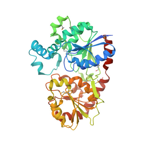
    3D0Q, 3D0R

  • PubMed Abstract: 

    The enediyne antibiotic calicheamicin (CLM) gamma(1)(I) is a prominent antitumor agent that is targeted to DNA by a novel aryltetrasaccharide comprised of an aromatic unit and four unusual carbohydrates. Herein we report the heterologous expression and the biochemical characterization of the two "internal" glycosyltransferases CalG3 and CalG2 and the structural elucidation of an enediyne glycosyltransferase (CalG3). In conjunction with the previous characterization of the "external" CLM GTs CalG1 and CalG4, this study completes the functional assignment of all four CLM GTs, extends the utility of enediyne GT-catalyzed reaction reversibility, and presents conclusive evidence of a sequential glycosylation pathway in CLM biosynthesis. This work also reveals the common GT-B structural fold can now be extended to include enediyne GTs.


  • Organizational Affiliation
    • Guangdong Key Laboratory of Marine Materia Medica, South China Sea Institute of Oceanology, Chinese Academy of Sciences, 164 Xingang West Road, Guangzhou, China.

Macromolecule Content 

  • Total Structure Weight: 87.19 kDa 
  • Atom Count: 5,787 
  • Modeled Residue Count: 754 
  • Deposited Residue Count: 796 
  • Unique protein chains: 1

Macromolecules

Find similar proteins by:|  3D Structure
Entity ID: 1
MoleculeChains  Sequence LengthOrganismDetailsImage
Protein CalG3
A, B
398Micromonospora echinosporaMutation(s): 0 
Gene Names: calG3
UniProt
Find proteins for Q8KND7 (Micromonospora echinospora)
Explore Q8KND7 
Go to UniProtKB:  Q8KND7
Entity Groups
Sequence Clusters30% Identity50% Identity70% Identity90% Identity95% Identity100% Identity
UniProt GroupQ8KND7
Sequence Annotations
Expand
Reference Sequence

Small Molecules

Modified Residues  1 Unique
IDChains TypeFormula2D DiagramParent
MSE
Query on MSE
A, B
L-PEPTIDE LINKINGC5 H11 N O2 SeMET

Experimental Data & Validation

Experimental Data

  • Method: X-RAY DIFFRACTION
  • Resolution: 2.79 Å
  • R-Value Free:  0.243 (Depositor), 0.236 (DCC) 
  • R-Value Work:  0.187 (Depositor), 0.193 (DCC) 
  • R-Value Observed: 0.190 (Depositor) 
Space Group: I 2 2 2
Unit Cell:
Length ( Å )Angle ( ˚ )
a = 106.654α = 90
b = 119.267β = 90
c = 155.942γ = 90
Software Package:
Software NamePurpose
DENZOdata reduction
SCALEPACKdata scaling
SHARPphasing
DMphasing
REFMACrefinement
PDB_EXTRACTdata extraction
MAR345data collection

Structure Validation

View Full Validation Report


Ligand Structure Quality Assessment 


Entry History 

Deposition Data

Revision History  (Full details and data files)

  • Version 1.0: 2008-06-24
    Type: Initial release
  • Version 1.1: 2011-07-13
    Changes: Advisory, Version format compliance
  • Version 1.2: 2017-10-25
    Changes: Refinement description
  • Version 1.3: 2024-11-06
    Changes: Data collection, Database references, Derived calculations, Refinement description, Structure summary