3D25 | pdb_00003d25

Crystal structure of HA-1 minor histocompatibility antigen bound to human class I MHC HLA-A2


Experimental Data Snapshot

  • Method: X-RAY DIFFRACTION
  • Resolution: 1.30 Å
  • R-Value Free: 
    0.204 (Depositor), 0.221 (DCC) 
  • R-Value Work: 
    0.204 (DCC) 
  • R-Value Observed: 
    0.169 (Depositor) 

Starting Model: experimental
View more details

wwPDB Validation 3D Report Full Report

Validation slider image for 3D25

This is version 1.3 of the entry. See complete history

Literature

Secondary anchor polymorphism in the HA-1 minor histocompatibility antigen critically affects MHC stability and TCR recognition

Nicholls, S.Piper, K.P.Mohammed, F.Dafforn, T.R.Tenzer, S.Salim, M.Mahendra, P.Craddock, C.van Endert, P.Schild, H.Cobbold, M.Engelhard, V.H.Moss, P.A.Willcox, B.E.

(2009) Proc Natl Acad Sci U S A 106: 3889-3894

  • DOI: https://doi.org/10.1073/pnas.0900411106
  • Primary Citation Related Structures: 
    3D25

  • PubMed Abstract: 

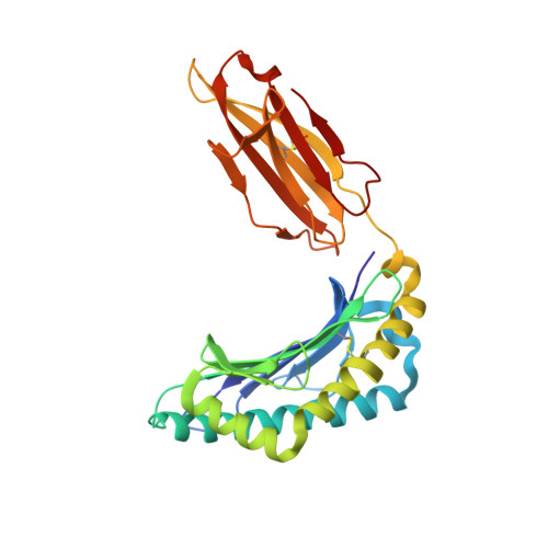
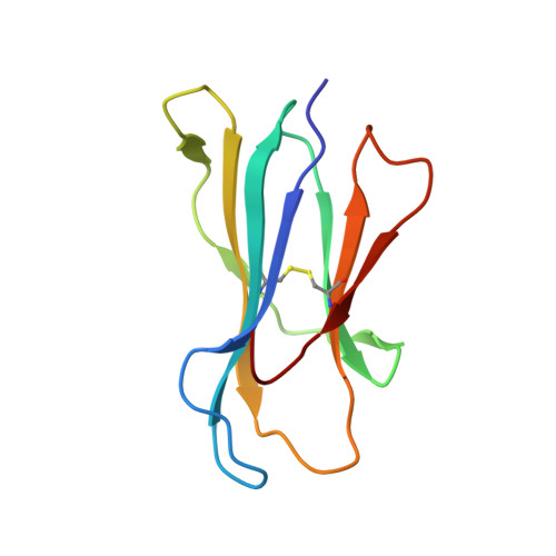
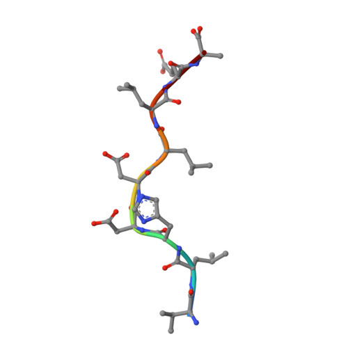
    T cell recognition of minor histocompatibility antigens (mHags) underlies allogeneic immune responses that mediate graft-versus-host disease and the graft-versus-leukemia effect following stem cell transplantation. Many mHags derive from single amino acid polymorphisms in MHC-restricted epitopes, but our understanding of the molecular mechanisms governing mHag immunogenicity and recognition is incomplete. Here we examined antigenic presentation and T-cell recognition of HA-1, a prototypic autosomal mHag derived from single nucleotide dimorphism (HA-1(H) versus HA-1(R)) in the HMHA1 gene. The HA-1(H) peptide is restricted by HLA-A2 and is immunogenic in HA-1(R/R) into HA-1(H) transplants, while HA-1(R) has been suggested to be a "null allele" in terms of T cell reactivity. We found that proteasomal cleavage and TAP transport of the 2 peptides is similar and that both variants can bind to MHC. However, the His>Arg change substantially decreases the stability and affinity of HLA-A2 association, consistent with the reduced immunogenicity of the HA-1(R) variant. To understand these findings, we determined the structure of an HLA-A2-HA-1(H) complex to 1.3A resolution. Whereas His-3 is accommodated comfortably in the D pocket, incorporation of the lengthy Arg-3 is predicted to require local conformational changes. Moreover, a soluble TCR generated from HA-1(H)-specific T-cells bound HA-1(H) peptide with moderate affinity but failed to bind HA-1(R), indicating complete discrimination of HA-1 variants at the level of TCR/MHC interaction. Our results define the molecular mechanisms governing immunogenicity of HA-1, and highlight how single amino acid polymorphisms in mHags can critically affect both MHC association and TCR recognition.


  • Organizational Affiliation
    • Cancer Research UK Institute for Cancer Studies, Edgbaston, Birmingham B15 2TT, United Kingdom.

Macromolecule Content 

  • Total Structure Weight: 44.39 kDa 
  • Atom Count: 3,508 
  • Modeled Residue Count: 381 
  • Deposited Residue Count: 381 
  • Unique protein chains: 3

Macromolecules

Find similar proteins by:|  3D Structure
Entity ID: 1
MoleculeChains  Sequence LengthOrganismDetailsImage
HLA class I histocompatibility antigen, A-2 alpha chain274Homo sapiensMutation(s): 0 
Gene Names: HLA-AHLAA
UniProt & NIH Common Fund Data Resources
Find proteins for P04439 (Homo sapiens)
Explore P04439 
Go to UniProtKB:  P04439
PHAROS:  P04439
GTEx:  ENSG00000206503 
Entity Groups
Sequence Clusters30% Identity50% Identity70% Identity90% Identity95% Identity100% Identity
UniProt GroupP04439
Sequence Annotations
Expand
Reference Sequence
Find similar proteins by:|  3D Structure
Entity ID: 2
MoleculeChains  Sequence LengthOrganismDetailsImage
Beta-2-microglobulin98Homo sapiensMutation(s): 0 
Gene Names: B2MCDABP0092HDCMA22P
UniProt & NIH Common Fund Data Resources
Find proteins for P61769 (Homo sapiens)
Explore P61769 
Go to UniProtKB:  P61769
PHAROS:  P61769
GTEx:  ENSG00000166710 
Entity Groups
Sequence Clusters30% Identity50% Identity70% Identity90% Identity95% Identity100% Identity
UniProt GroupP61769
Sequence Annotations
Expand
Reference Sequence
Find similar proteins by:  Sequence   |   3D Structure  
Entity ID: 3
MoleculeChains  Sequence LengthOrganismDetailsImage
Nonameric peptide from HA-19N/AMutation(s): 0 
UniProt & NIH Common Fund Data Resources
Find proteins for Q92619 (Homo sapiens)
Explore Q92619 
Go to UniProtKB:  Q92619
PHAROS:  Q92619
GTEx:  ENSG00000180448 
Entity Groups
UniProt GroupQ92619
Sequence Annotations
Expand
Reference Sequence

Experimental Data & Validation

Experimental Data

  • Method: X-RAY DIFFRACTION
  • Resolution: 1.30 Å
  • R-Value Free:  0.204 (Depositor), 0.221 (DCC) 
  • R-Value Work:  0.204 (DCC) 
  • R-Value Observed: 0.169 (Depositor) 
Space Group: P 1 21 1
Unit Cell:
Length ( Å )Angle ( ˚ )
a = 53.17α = 90
b = 80.98β = 112.32
c = 56.26γ = 90
Software Package:
Software NamePurpose
SCALAdata processing
SHELXrefinement
PDB_EXTRACTdata extraction
MOSFLMdata reduction
SCALAdata scaling
CNSphasing
SHELXL-97refinement

Structure Validation

View Full Validation Report


Entry History 

Revision History  (Full details and data files)

  • Version 1.0: 2009-02-10
    Type: Initial release
  • Version 1.1: 2011-07-13
    Changes: Version format compliance
  • Version 1.2: 2023-08-30
    Changes: Data collection, Database references, Refinement description
  • Version 1.3: 2024-10-09
    Changes: Structure summary