3D2U | pdb_00003d2u

Structure of UL18, a Peptide-Binding Viral MHC Mimic, Bound to a Host Inhibitory Receptor


Experimental Data Snapshot

  • Method: X-RAY DIFFRACTION
  • Resolution: 2.21 Å
  • R-Value Free: 
    0.259 (Depositor) 
  • R-Value Work: 
    0.241 (Depositor), 0.236 (DCC) 

Starting Model: experimental
View more details

wwPDB Validation 3D Report Full Report

Validation slider image for 3D2U

Ligand Structure Quality Assessment 


This is version 1.7 of the entry. See complete history

Literature

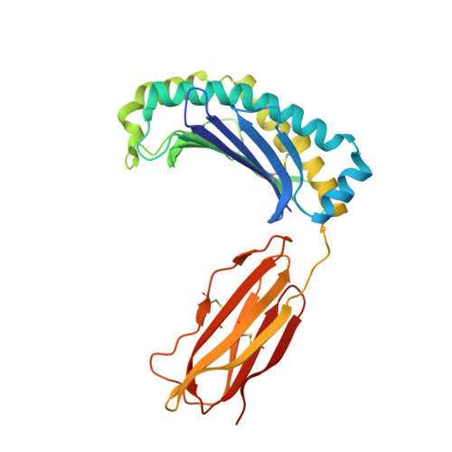
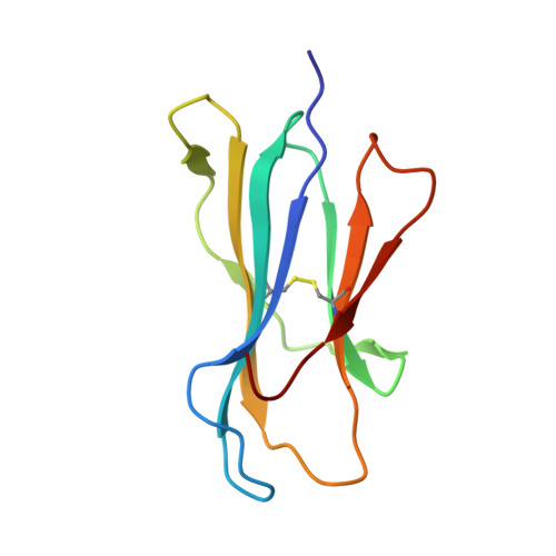
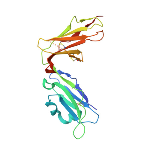
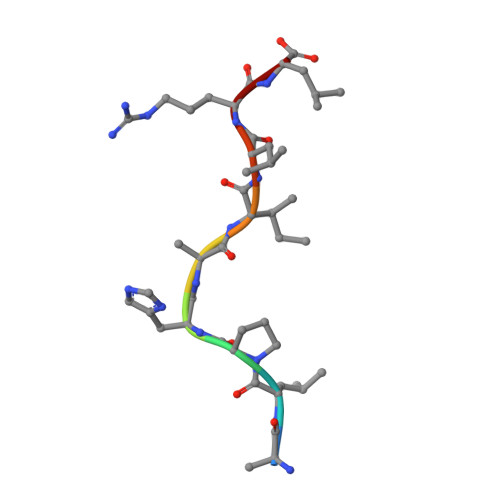
Structure of UL18, a peptide-binding viral MHC mimic, bound to a host inhibitory receptor

Yang, Z.Bjorkman, P.J.

(2008) Proc Natl Acad Sci U S A 105: 10095-10100

  • DOI: https://doi.org/10.1073/pnas.0804551105
  • Primary Citation Related Structures: 
    3D2U

  • PubMed Abstract: 

    UL18 is a human cytomegalovirus class I MHC (MHCI) homolog that binds the host inhibitory receptor LIR-1 and the only known viral MHC homolog that presents peptides. The 2.2-A structure of a LIR-1/UL18/peptide complex reveals increased contacts and optimal surface complementarity in the LIR-1/UL18 interface compared with LIR/MHCI interfaces, resulting in a >1,000-fold higher affinity. Despite sharing only approximately 25% sequence identity, UL18's structure and peptide binding are surprisingly similar to host MHCI. The crystal structure suggests that most of the UL18 surface, except where LIR-1 and the host-derived light chain bind, is covered by carbohydrates attached to 13 potential N-glycosylation sites, thereby preventing access to bound peptide and association with most MHCI-binding proteins. The LIR-1/UL18 structure demonstrates how a viral protein evolves from its host ancestor to impede unwanted interactions while preserving and improving its receptor-binding site.


  • Organizational Affiliation
    • Division of Biology 114-96 and Howard Hughes Medical Institute, California Institute of Technology, Pasadena, CA 91125, USA.

Macromolecule Content 

  • Total Structure Weight: 136.35 kDa 
  • Atom Count: 9,681 
  • Modeled Residue Count: 1,143 
  • Deposited Residue Count: 1,174 
  • Unique protein chains: 4

Macromolecules

Find similar proteins by:|  3D Structure
Entity ID: 1
MoleculeChains  Sequence LengthOrganismDetailsImage
UL18 proteinA,
D [auth E]
281Human betaherpesvirus 5Mutation(s): 5 
UniProt
Find proteins for P08560 (Human cytomegalovirus (strain AD169))
Explore P08560 
Go to UniProtKB:  P08560
Entity Groups
Sequence Clusters30% Identity50% Identity70% Identity90% Identity95% Identity100% Identity
UniProt GroupP08560
Sequence Annotations
Expand
Reference Sequence
Find similar proteins by:|  3D Structure
Entity ID: 2
MoleculeChains  Sequence LengthOrganismDetailsImage
Beta-2-microglobulinB,
E [auth F]
99Homo sapiensMutation(s): 3 
UniProt & NIH Common Fund Data Resources
Find proteins for P61769 (Homo sapiens)
Explore P61769 
Go to UniProtKB:  P61769
PHAROS:  P61769
GTEx:  ENSG00000166710 
Entity Groups
Sequence Clusters30% Identity50% Identity70% Identity90% Identity95% Identity100% Identity
UniProt GroupP61769
Sequence Annotations
Expand
Reference Sequence
Find similar proteins by:|  3D Structure
Entity ID: 3
MoleculeChains  Sequence LengthOrganismDetailsImage
Leukocyte immunoglobulin-like receptor subfamily B member 1C [auth D],
F [auth H]
198Homo sapiensMutation(s): 3 
UniProt & NIH Common Fund Data Resources
Find proteins for Q8NHL6 (Homo sapiens)
Explore Q8NHL6 
Go to UniProtKB:  Q8NHL6
PHAROS:  Q8NHL6
Entity Groups
Sequence Clusters30% Identity50% Identity70% Identity90% Identity95% Identity100% Identity
UniProt GroupQ8NHL6
Sequence Annotations
Expand
Reference Sequence
Find similar proteins by:  Sequence   |   3D Structure  
Entity ID: 4
MoleculeChains  Sequence LengthOrganismDetailsImage
ActinG,
H [auth C]
9Homo sapiensMutation(s): 5 
EC: 3.6.4
UniProt & NIH Common Fund Data Resources
Find proteins for P60709 (Homo sapiens)
Explore P60709 
Go to UniProtKB:  P60709
PHAROS:  P60709
GTEx:  ENSG00000075624 
Entity Groups
UniProt GroupP60709
Sequence Annotations
Expand
Reference Sequence

Small Molecules

Ligands 4 Unique
IDChains Name / Formula / InChI Key2D Diagram3D Interactions
NAG

Query on NAG


Download:Ideal Coordinates CCD File
I [auth A]
J [auth A]
N [auth A]
O [auth A]
P [auth A]
I [auth A],
J [auth A],
N [auth A],
O [auth A],
P [auth A],
Q [auth A],
R [auth A],
S [auth E],
T [auth E],
U [auth E],
V [auth E],
W [auth E],
X [auth E]
2-acetamido-2-deoxy-beta-D-glucopyranose
C8 H15 N O6
OVRNDRQMDRJTHS-FMDGEEDCSA-N
BMA

Query on BMA


Download:Ideal Coordinates CCD File
L [auth A]beta-D-mannopyranose
C6 H12 O6
WQZGKKKJIJFFOK-RWOPYEJCSA-N
MAN

Query on MAN


Download:Ideal Coordinates CCD File
K [auth A]alpha-D-mannopyranose
C6 H12 O6
WQZGKKKJIJFFOK-PQMKYFCFSA-N
FUC

Query on FUC


Download:Ideal Coordinates CCD File
M [auth A]alpha-L-fucopyranose
C6 H12 O5
SHZGCJCMOBCMKK-SXUWKVJYSA-N

Experimental Data & Validation

Experimental Data

  • Method: X-RAY DIFFRACTION
  • Resolution: 2.21 Å
  • R-Value Free:  0.259 (Depositor) 
  • R-Value Work:  0.241 (Depositor), 0.236 (DCC) 
Space Group: P 1 21 1
Unit Cell:
Length ( Å )Angle ( ˚ )
a = 52.366α = 90
b = 98.11β = 98.46
c = 172.147γ = 90
Software Package:
Software NamePurpose
DENZOdata reduction
SCALEPACKdata scaling
PHASERphasing
DMphasing
CNSrefinement
PDB_EXTRACTdata extraction
Blu-Icedata collection

Structure Validation

View Full Validation Report


Ligand Structure Quality Assessment 


Entry History 

Deposition Data

Revision History  (Full details and data files)

  • Version 1.0: 2008-07-08
    Type: Initial release
  • Version 1.1: 2011-07-13
    Changes: Non-polymer description, Version format compliance
  • Version 1.2: 2017-10-25
    Changes: Advisory, Refinement description
  • Version 1.3: 2019-12-25
    Changes: Data collection, Database references, Derived calculations, Refinement description, Source and taxonomy, Structure summary
  • Version 1.4: 2020-07-29
    Type: Remediation
    Reason: Carbohydrate remediation
    Changes: Data collection, Derived calculations, Structure summary
  • Version 1.5: 2021-10-20
    Changes: Advisory, Database references, Structure summary
  • Version 1.6: 2023-08-30
    Changes: Data collection, Refinement description
  • Version 1.7: 2024-11-20
    Changes: Structure summary