3D7Z | pdb_00003d7z

Crystal Structure of P38 Kinase in Complex with a biphenyl amide inhibitor


Experimental Data Snapshot

  • Method: X-RAY DIFFRACTION
  • Resolution: 2.10 Å
  • R-Value Free: 
    0.227 (Depositor), 0.229 (DCC) 
  • R-Value Work: 
    0.174 (Depositor), 0.177 (DCC) 
  • R-Value Observed: 
    0.177 (Depositor) 

Starting Model: experimental
View more details

wwPDB Validation 3D Report Full Report

Validation slider image for 3D7Z

Ligand Structure Quality Assessment 


This is version 1.5 of the entry. See complete history

Literature

Biphenyl amide p38 kinase inhibitors 3: Improvement of cellular and in vivo activity.

Angell, R.Aston, N.M.Bamborough, P.Buckton, J.B.Cockerill, S.deBoeck, S.J.Edwards, C.D.Holmes, D.S.Jones, K.L.Laine, D.I.Patel, S.Smee, P.A.Smith, K.J.Somers, D.O.Walker, A.L.

(2008) Bioorg Med Chem Lett 18: 4428-4432

  • DOI: https://doi.org/10.1016/j.bmcl.2008.06.048
  • Primary Citation Related Structures: 
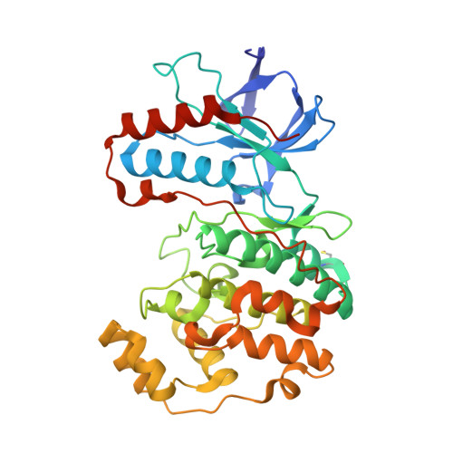
    3D7Z

  • PubMed Abstract: 

    The biphenyl amides (BPAs) are a novel series of p38alpha MAP kinase inhibitor. The optimisation of the series to give compounds that are potent in an in vivo disease model is discussed. SAR is presented and rationalised with reference to the crystallographic binding mode.


  • Organizational Affiliation
    • GlaxoSmithKline R&D, Medicines Research Centre, Gunnels Wood Road, Stevenage, Hertfordshire SG1 2NY, UK.

Macromolecule Content 

  • Total Structure Weight: 41.9 kDa 
  • Atom Count: 3,225 
  • Modeled Residue Count: 349 
  • Deposited Residue Count: 360 
  • Unique protein chains: 1

Macromolecules

Find similar proteins by:|  3D Structure
Entity ID: 1
MoleculeChains  Sequence LengthOrganismDetailsImage
Mitogen-activated protein kinase 14360Homo sapiensMutation(s): 0 
Gene Names: MAPK14CSBPCSBP1CSBP2CSPB1MXI2
EC: 2.7.11.24
UniProt & NIH Common Fund Data Resources
Find proteins for Q16539 (Homo sapiens)
Explore Q16539 
Go to UniProtKB:  Q16539
PHAROS:  Q16539
GTEx:  ENSG00000112062 
Entity Groups
Sequence Clusters30% Identity50% Identity70% Identity90% Identity95% Identity100% Identity
UniProt GroupQ16539
Sequence Annotations
Expand
Reference Sequence

Small Molecules

Modified Residues  1 Unique
IDChains TypeFormula2D DiagramParent
CSO
Query on CSO
A
L-PEPTIDE LINKINGC3 H7 N O3 SCYS

Experimental Data & Validation

Experimental Data

  • Method: X-RAY DIFFRACTION
  • Resolution: 2.10 Å
  • R-Value Free:  0.227 (Depositor), 0.229 (DCC) 
  • R-Value Work:  0.174 (Depositor), 0.177 (DCC) 
  • R-Value Observed: 0.177 (Depositor) 
Space Group: P 21 21 21
Unit Cell:
Length ( Å )Angle ( ˚ )
a = 45.63α = 90
b = 86.103β = 90
c = 124.811γ = 90
Software Package:
Software NamePurpose
DENZOdata reduction
SCALEPACKdata scaling
REFMACrefinement
PDB_EXTRACTdata extraction
REFMACphasing

Structure Validation

View Full Validation Report


Ligand Structure Quality Assessment 


Entry History 

Deposition Data

Revision History  (Full details and data files)

  • Version 1.0: 2008-07-22
    Type: Initial release
  • Version 1.1: 2011-07-13
    Changes: Advisory, Refinement description, Version format compliance
  • Version 1.2: 2017-10-25
    Changes: Refinement description
  • Version 1.3: 2023-08-30
    Changes: Data collection, Database references, Derived calculations, Refinement description
  • Version 1.4: 2023-11-15
    Changes: Data collection
  • Version 1.5: 2024-11-13
    Changes: Structure summary