3D83 | pdb_00003d83

Crystal structure of P38 kinase in complex with a biphenyl amide inhibitor


Experimental Data Snapshot

  • Method: X-RAY DIFFRACTION
  • Resolution: 1.90 Å
  • R-Value Free: 
    0.218 (Depositor), 0.219 (DCC) 
  • R-Value Work: 
    0.175 (Depositor), 0.177 (DCC) 
  • R-Value Observed: 
    0.177 (Depositor) 

Starting Model: experimental
View more details

wwPDB Validation 3D Report Full Report

Validation slider image for 3D83

Ligand Structure Quality Assessment 


This is version 1.5 of the entry. See complete history

Literature

Biphenyl amide p38 kinase inhibitors 4: DFG-in and DFG-out binding modes.

Angell, R.M.Angell, T.D.Bamborough, P.Bamford, M.J.Chung, C.W.Cockerill, S.G.Flack, S.S.Jones, K.L.Laine, D.I.Longstaff, T.Ludbrook, S.Pearson, R.Smith, K.J.Smee, P.A.Somers, D.O.Walker, A.L.

(2008) Bioorg Med Chem Lett 18: 4433-4437

  • DOI: https://doi.org/10.1016/j.bmcl.2008.06.028
  • Primary Citation Related Structures: 
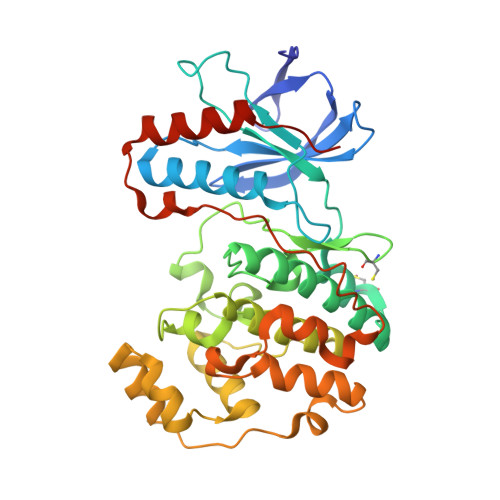
    3D83

  • PubMed Abstract: 

    The biphenyl amides (BPAs) are a series of p38alpha MAP kinase inhibitors. Compounds are able to bind to the kinase in either the DFG-in or DFG-out conformation, depending on substituents. X-ray, binding, kinetic and cellular data are shown, providing the most detailed comparison to date between potent compounds from the same chemical series that bind to different p38alpha conformations. DFG-out-binding compounds could be made more potent than DFG-in-binding compounds by increasing their size. Unexpectedly, compounds that bound to the DGF-out conformation showed diminished selectivity. The kinetics of binding to the isolated enzyme and the effects of compounds on cells were largely unaffected by the kinase conformation bound.


  • Organizational Affiliation
    • GlaxoSmithKline R&D, Medicines Research Centre, Gunnels Wood Road, Stevenage, Hertfordshire SG1 2NY, UK.

Macromolecule Content 

  • Total Structure Weight: 41.94 kDa 
  • Atom Count: 3,291 
  • Modeled Residue Count: 349 
  • Deposited Residue Count: 360 
  • Unique protein chains: 1

Macromolecules

Find similar proteins by:|  3D Structure
Entity ID: 1
MoleculeChains  Sequence LengthOrganismDetailsImage
Mitogen-activated protein kinase 14360Homo sapiensMutation(s): 0 
Gene Names: MAPK14CSBPCSBP1CSBP2CSPB1MXI2
EC: 2.7.11.24
UniProt & NIH Common Fund Data Resources
Find proteins for Q16539 (Homo sapiens)
Explore Q16539 
Go to UniProtKB:  Q16539
PHAROS:  Q16539
GTEx:  ENSG00000112062 
Entity Groups
Sequence Clusters30% Identity50% Identity70% Identity90% Identity95% Identity100% Identity
UniProt GroupQ16539
Sequence Annotations
Expand
Reference Sequence

Small Molecules

Ligands 2 Unique
IDChains Name / Formula / InChI Key2D Diagram3D Interactions
GK6

Query on GK6


Download:Ideal Coordinates CCD File
B [auth A]N-{4'-[(cyclopropylmethyl)carbamoyl]-6-methylbiphenyl-3-yl}-2-morpholin-4-ylpyridine-4-carboxamide
C28 H30 N4 O3
BOQNSWFZUHQNRG-UHFFFAOYSA-N
GOL

Query on GOL


Download:Ideal Coordinates CCD File
C [auth A]GLYCEROL
C3 H8 O3
PEDCQBHIVMGVHV-UHFFFAOYSA-N
Modified Residues  1 Unique
IDChains TypeFormula2D DiagramParent
CSO
Query on CSO
A
L-PEPTIDE LINKINGC3 H7 N O3 SCYS

Experimental Data & Validation

Experimental Data

  • Method: X-RAY DIFFRACTION
  • Resolution: 1.90 Å
  • R-Value Free:  0.218 (Depositor), 0.219 (DCC) 
  • R-Value Work:  0.175 (Depositor), 0.177 (DCC) 
  • R-Value Observed: 0.177 (Depositor) 
Space Group: P 21 21 21
Unit Cell:
Length ( Å )Angle ( ˚ )
a = 46.24α = 90
b = 86.523β = 90
c = 127.108γ = 90
Software Package:
Software NamePurpose
DENZOdata reduction
SCALEPACKdata scaling
REFMACrefinement
PDB_EXTRACTdata extraction
ADSCdata collection
REFMACphasing

Structure Validation

View Full Validation Report


Ligand Structure Quality Assessment 


Entry History 

Deposition Data

  • Released Date: 2008-07-22 
  • Deposition Author(s): Somers, D.O.

Revision History  (Full details and data files)

  • Version 1.0: 2008-07-22
    Type: Initial release
  • Version 1.1: 2011-07-13
    Changes: Advisory, Refinement description, Version format compliance
  • Version 1.2: 2017-10-25
    Changes: Refinement description
  • Version 1.3: 2023-08-30
    Changes: Data collection, Database references, Derived calculations, Refinement description
  • Version 1.4: 2023-11-15
    Changes: Data collection
  • Version 1.5: 2024-10-30
    Changes: Structure summary