3DVM | pdb_00003dvm

Crystal Structure of Ca2+/CaM-CaV2.1 IQ domain complex


Experimental Data Snapshot

  • Method: X-RAY DIFFRACTION
  • Resolution: 2.60 Å
  • R-Value Free: 
    0.320 (Depositor), 0.314 (DCC) 
  • R-Value Work: 
    0.276 (Depositor), 0.274 (DCC) 
  • R-Value Observed: 
    0.278 (Depositor) 

wwPDB Validation 3D Report Full Report

Validation slider image for 3DVM

This is version 1.3 of the entry. See complete history

Literature

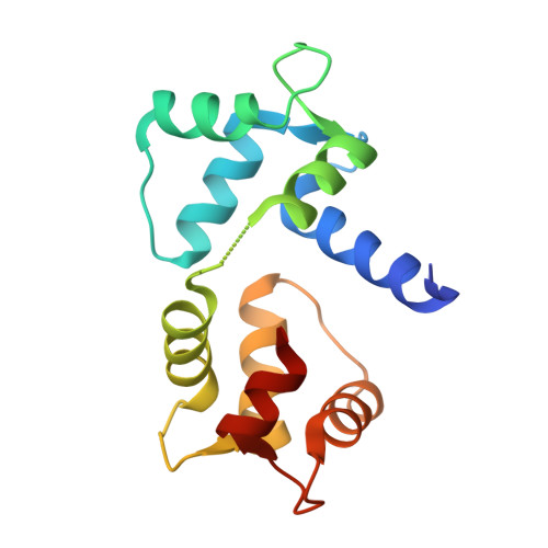
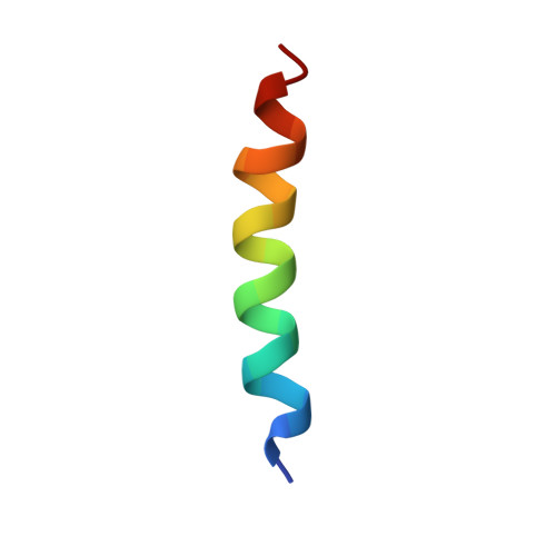
Structures of Ca(V)2 Ca(2+)/CaM-IQ Domain Complexes Reveal Binding Modes that Underlie Calcium-Dependent Inactivation and Facilitation.

Kim, E.Y.Rumpf, C.H.Fujiwara, Y.Cooley, E.S.Van Petegem, F.Minor, D.L.

(2008) Structure 16: 1455-1467

  • DOI: https://doi.org/10.1016/j.str.2008.07.010
  • Primary Citation Related Structures: 
    3DVE, 3DVJ, 3DVK, 3DVM

  • PubMed Abstract: 

    Calcium influx drives two opposing voltage-activated calcium channel (Ca(V)) self-modulatory processes: calcium-dependent inactivation (CDI) and calcium-dependent facilitation (CDF). Specific Ca(2+)/calmodulin (Ca(2+)/CaM) lobes produce CDI and CDF through interactions with the Ca(V)alpha(1) subunit IQ domain. Curiously, Ca(2+)/CaM lobe modulation polarity appears inverted between Ca(V)1s and Ca(V)2s. Here, we present crystal structures of Ca(V)2.1, Ca(V)2.2, and Ca(V)2.3 Ca(2+)/CaM-IQ domain complexes. All display binding orientations opposite to Ca(V)1.2 with a physical reversal of the CaM lobe positions relative to the IQ alpha-helix. Titration calorimetry reveals lobe competition for a high-affinity site common to Ca(V)1 and Ca(V)2 IQ domains that is occupied by the CDI lobe in the structures. Electrophysiological experiments demonstrate that the N-terminal Ca(V)2 Ca(2+)/C-lobe anchors affect CDF. Together, the data unveil the remarkable structural plasticity at the heart of Ca(V) feedback modulation and indicate that Ca(V)1 and Ca(V)2 IQ domains bear a dedicated CDF site that exchanges Ca(2+)/CaM lobe occupants.


  • Organizational Affiliation
    • Cardiovascular Research Institute, Department of Biochemistry and Biophysics, California Institute for Quantitative Biosciences, University of California, San Francisco, CA 94158-2330, USA.

Macromolecule Content 

  • Total Structure Weight: 19.57 kDa 
  • Atom Count: 1,188 
  • Modeled Residue Count: 162 
  • Deposited Residue Count: 170 
  • Unique protein chains: 2

Macromolecules

Find similar proteins by:|  3D Structure
Entity ID: 1
MoleculeChains  Sequence LengthOrganismDetailsImage
Calmodulin148Homo sapiensMutation(s): 0 
Gene Names: 
CALM1CALMCAMCAM1CALM2CAM2CAMBCALM3CALML2CAM3...
CALM1CALMCAMCAM1CALM2CAM2CAMBCALM3CALML2CAM3CAMCCAMIII

UniProt & NIH Common Fund Data Resources
Find proteins for P0DP23 (Homo sapiens)
Explore P0DP23 
Go to UniProtKB:  P0DP23
PHAROS:  P0DP23
GTEx:  ENSG00000198668 
Entity Groups
Sequence Clusters30% Identity50% Identity70% Identity90% Identity95% Identity100% Identity
UniProt GroupP0DP23
Sequence Annotations
Expand
Reference Sequence
Find similar proteins by:  Sequence   |   3D Structure  
Entity ID: 2
MoleculeChains  Sequence LengthOrganismDetailsImage
Voltage-dependent P/Q-type calcium channel subunit alpha-1A22Oryctolagus cuniculusMutation(s): 0 
Gene Names: CACNA1ACACH4CACN3CACNL1A4
UniProt
Find proteins for P27884 (Oryctolagus cuniculus)
Explore P27884 
Go to UniProtKB:  P27884
Entity Groups
Sequence Clusters30% Identity50% Identity70% Identity90% Identity95% Identity100% Identity
UniProt GroupP27884
Sequence Annotations
Expand
Reference Sequence

Experimental Data & Validation

Experimental Data

  • Method: X-RAY DIFFRACTION
  • Resolution: 2.60 Å
  • R-Value Free:  0.320 (Depositor), 0.314 (DCC) 
  • R-Value Work:  0.276 (Depositor), 0.274 (DCC) 
  • R-Value Observed: 0.278 (Depositor) 
Space Group: P 61 2 2
Unit Cell:
Length ( Å )Angle ( ˚ )
a = 43.956α = 90
b = 43.956β = 90
c = 337.87γ = 120
Software Package:
Software NamePurpose
DENZOdata reduction
SCALEPACKdata scaling
SOLVEphasing
RESOLVEphasing
REFMACrefinement
PDB_EXTRACTdata extraction
HKL-2000data collection
HKL-2000data reduction
HKL-2000data scaling

Structure Validation

View Full Validation Report


Entry History 

Deposition Data

Revision History  (Full details and data files)

  • Version 1.0: 2008-11-04
    Type: Initial release
  • Version 1.1: 2011-07-13
    Changes: Advisory, Version format compliance
  • Version 1.2: 2017-10-25
    Changes: Refinement description
  • Version 1.3: 2024-02-21
    Changes: Data collection, Database references, Derived calculations