3E4A

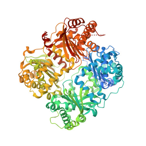
Human IDE-inhibitor complex at 2.6 angstrom resolution


Experimental Data Snapshot

  • Method: X-RAY DIFFRACTION
  • Resolution: 2.60 Å
  • R-Value Free: 0.225 
  • R-Value Work: 0.167 

Starting Model: experimental
View more details

wwPDB Validation   3D Report Full Report


Ligand Structure Quality Assessment 


This is version 1.4 of the entry. See complete history


Literature

Designed inhibitors of insulin-degrading enzyme regulate the catabolism and activity of insulin.

Leissring, M.A.Malito, E.Hedouin, S.Reinstatler, L.Sahara, T.Abdul-Hay, S.O.Choudhry, S.Maharvi, G.M.Fauq, A.H.Huzarska, M.May, P.S.Choi, S.Logan, T.P.Turk, B.E.Cantley, L.C.Manolopoulou, M.Tang, W.J.Stein, R.L.Cuny, G.D.Selkoe, D.J.

(2010) PLoS One 5: e10504-e10504

  • DOI: https://doi.org/10.1371/journal.pone.0010504
  • Primary Citation of Related Structures:  
    3E4A

  • PubMed Abstract: 

    Insulin is a vital peptide hormone that is a central regulator of glucose homeostasis, and impairments in insulin signaling cause diabetes mellitus. In principle, it should be possible to enhance the activity of insulin by inhibiting its catabolism, which is mediated primarily by insulin-degrading enzyme (IDE), a structurally and evolutionarily distinctive zinc-metalloprotease. Despite interest in pharmacological inhibition of IDE as an attractive anti-diabetic approach dating to the 1950s, potent and selective inhibitors of IDE have not yet emerged. We used a rational design approach based on analysis of combinatorial peptide mixtures and focused compound libraries to develop novel peptide hydroxamic acid inhibitors of IDE. The resulting compounds are approximately 10(6) times more potent than existing inhibitors, non-toxic, and surprisingly selective for IDE vis-à-vis conventional zinc-metalloproteases. Crystallographic analysis of an IDE-inhibitor complex reveals a novel mode of inhibition based on stabilization of IDE's "closed," inactive conformation. We show further that pharmacological inhibition of IDE potentiates insulin signaling by a mechanism involving reduced catabolism of internalized insulin. The inhibitors we describe are the first to potently and selectively inhibit IDE or indeed any member of this atypical zinc-metalloprotease superfamily. The distinctive structure of IDE's active site, and the mode of action of our inhibitors, suggests that it may be possible to develop inhibitors that cross-react minimally with conventional zinc-metalloproteases. Significantly, our results reveal that insulin signaling is normally regulated by IDE activity not only extracellularly but also within cells, supporting the longstanding view that IDE inhibitors could hold therapeutic value for the treatment of diabetes.


  • Organizational Affiliation

    Department of Neuroscience, Mayo Clinic Florida, Jacksonville, Florida, United States of America. Leissring@mayo.edu


Macromolecules
Find similar proteins by:  (by identity cutoff)  |  3D Structure
Entity ID: 1
MoleculeChains Sequence LengthOrganismDetailsImage
Insulin-degrading enzyme
A, B
1,019Homo sapiensMutation(s): 20 
Gene Names: IDE
EC: 3.4.24.56
UniProt & NIH Common Fund Data Resources
Find proteins for P14735 (Homo sapiens)
Explore P14735 
Go to UniProtKB:  P14735
PHAROS:  P14735
GTEx:  ENSG00000119912 
Entity Groups  
Sequence Clusters30% Identity50% Identity70% Identity90% Identity95% Identity100% Identity
UniProt GroupP14735
Sequence Annotations
Expand
  • Reference Sequence

Find similar proteins by:  Sequence   |   3D Structure  

Entity ID: 2
MoleculeChains Sequence LengthOrganismDetailsImage
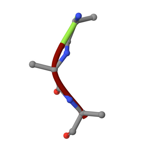
HYDROXAMATE PEPTIDE II1C [auth F],
D [auth G]
3synthetic constructMutation(s): 0 
Sequence Annotations
Expand
  • Reference Sequence
Small Molecules
Ligands 4 Unique
IDChains Name / Formula / InChI Key2D Diagram3D Interactions
QIX
Query on QIX

Download Ideal Coordinates CCD File 
F [auth A],
R [auth B]
N~2~-[(2R)-4-(HYDROXYAMINO)-2-(2-NAPHTHYLMETHYL)-4-OXOBUTANOYL]-L-ARGINYLGLYCYLGLYCINAMIDE
C25 H34 N8 O6
NINUSDCYEXCHSI-MOPGFXCFSA-N
DIO
Query on DIO

Download Ideal Coordinates CCD File 
G [auth A]
H [auth A]
S [auth B]
T [auth B]
U [auth B]
G [auth A],
H [auth A],
S [auth B],
T [auth B],
U [auth B],
V [auth B],
W [auth B]
1,4-DIETHYLENE DIOXIDE
C4 H8 O2
RYHBNJHYFVUHQT-UHFFFAOYSA-N
ZN
Query on ZN

Download Ideal Coordinates CCD File 
E [auth A],
Q [auth B]
ZINC ION
Zn
PTFCDOFLOPIGGS-UHFFFAOYSA-N
ACY
Query on ACY

Download Ideal Coordinates CCD File 
AA [auth B]
BA [auth B]
CA [auth B]
I [auth A]
J [auth A]
AA [auth B],
BA [auth B],
CA [auth B],
I [auth A],
J [auth A],
K [auth A],
L [auth A],
M [auth A],
N [auth A],
O [auth A],
P [auth A],
X [auth B],
Y [auth B],
Z [auth B]
ACETIC ACID
C2 H4 O2
QTBSBXVTEAMEQO-UHFFFAOYSA-N
Binding Affinity Annotations 
IDSourceBinding Affinity
QIX PDBBind:  3E4A Ki: 1.7 (nM) from 1 assay(s)
Experimental Data & Validation

Experimental Data

  • Method: X-RAY DIFFRACTION
  • Resolution: 2.60 Å
  • R-Value Free: 0.225 
  • R-Value Work: 0.167 
  • Space Group: P 65
Unit Cell:
Length ( Å )Angle ( ˚ )
a = 261.402α = 90
b = 261.402β = 90
c = 92.054γ = 120
Software Package:
Software NamePurpose
PHASERphasing
PHENIXrefinement
SBC-Collectdata collection
HKL-2000data reduction
SCALEPACKdata scaling

Structure Validation

View Full Validation Report


Ligand Structure Quality Assessment 


Entry History 

Deposition Data

Revision History  (Full details and data files)

  • Version 1.0: 2009-05-19
    Type: Initial release
  • Version 1.1: 2011-07-13
    Changes: Version format compliance
  • Version 1.2: 2017-10-25
    Changes: Refinement description, Source and taxonomy
  • Version 1.3: 2021-10-20
    Changes: Database references, Derived calculations
  • Version 1.4: 2023-08-30
    Changes: Data collection, Refinement description