3E59 | pdb_00003e59

Crystal structure of the PvcA (PA2254) protein from Pseudomonas aeruginosa


Experimental Data Snapshot

  • Method: X-RAY DIFFRACTION
  • Resolution: 2.10 Å
  • R-Value Free: 
    0.246 (Depositor), 0.245 (DCC) 
  • R-Value Work: 
    0.197 (Depositor), 0.199 (DCC) 
  • R-Value Observed: 
    0.200 (Depositor) 

wwPDB Validation 3D Report Full Report

Validation slider image for 3E59

This is version 1.2 of the entry. See complete history

Literature

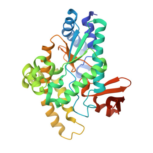
Three-dimensional structures of Pseudomonas aeruginosa PvcA and PvcB, two proteins involved in the synthesis of 2-isocyano-6,7-dihydroxycoumarin.

Drake, E.J.Gulick, A.M.

(2008) J Mol Biology 384: 193-205

  • DOI: https://doi.org/10.1016/j.jmb.2008.09.027
  • Primary Citation Related Structures: 
    3E59, 3EAT

  • PubMed Abstract: 

    The pvcABCD operon of Pseudomonas aeruginosa encodes four proteins (PA2254, PA2255, PA2256, and PA2257) that form a cluster that is responsible for the synthesis of a cyclized isocyano derivative of tyrosine. These proteins, which were identified originally as being responsible for a step in the maturation of the chromophore of the peptide siderophore pyoverdine, have been identified recently as belonging to a family of proteins that produce small organic isonitriles. We report that strains harboring a disruption in the pvcA or pvcB genes are able to grow in iron-depleted conditions and to produce pyoverdine. Additionally, we have determined the three-dimensional crystal structures of PvcA and PvcB. The structure of PvcA demonstrates a novel enzyme architecture that is built upon a Rossmann fold. We have analyzed the sequence conservation of enzymes within this family and identified six conserved motifs. These regions of the protein cluster around a putative active site cavity. The structure of the PvcB protein confirms it is a member of the Fe2+/alpha-ketoglutarate-dependent oxygenase family of enzymes. The active site of PvcB is compared to the structures of other family members and suggests that a conformational change to order several loops will accompany the binding of ligands.


  • Organizational Affiliation
    • Hauptman-Woodward Medical Research Institute, Department of Structural Biology, State University of New York at Buffalo, 700 Ellicott St, Buffalo, NY 14203-1102, USA.

Macromolecule Content 

  • Total Structure Weight: 150.38 kDa 
  • Atom Count: 10,092 
  • Modeled Residue Count: 1,232 
  • Deposited Residue Count: 1,320 
  • Unique protein chains: 1

Macromolecules

Find similar proteins by:|  3D Structure
Entity ID: 1
MoleculeChains  Sequence LengthOrganismDetailsImage
Pyoverdine biosynthesis protein PvcA
A, B, C, D
330Pseudomonas aeruginosaMutation(s): 0 
Gene Names: pvcAPA2254
EC: 4.1.99.24
UniProt
Find proteins for Q9I1L5 (Pseudomonas aeruginosa (strain ATCC 15692 / DSM 22644 / CIP 104116 / JCM 14847 / LMG 12228 / 1C / PRS 101 / PAO1))
Explore Q9I1L5 
Go to UniProtKB:  Q9I1L5
Entity Groups
Sequence Clusters30% Identity50% Identity70% Identity90% Identity95% Identity100% Identity
UniProt GroupQ9I1L5
Sequence Annotations
Expand
Reference Sequence

Small Molecules

Ligands 2 Unique
IDChains Name / Formula / InChI Key2D Diagram3D Interactions
PGE

Query on PGE


Download:Ideal Coordinates CCD File
H [auth B],
I [auth B]
TRIETHYLENE GLYCOL
C6 H14 O4
ZIBGPFATKBEMQZ-UHFFFAOYSA-N
PO4

Query on PO4


Download:Ideal Coordinates CCD File
E [auth A]
F [auth A]
G [auth B]
J [auth C]
K [auth D]
E [auth A],
F [auth A],
G [auth B],
J [auth C],
K [auth D],
L [auth D]
PHOSPHATE ION
O4 P
NBIIXXVUZAFLBC-UHFFFAOYSA-K

Experimental Data & Validation

Experimental Data

  • Method: X-RAY DIFFRACTION
  • Resolution: 2.10 Å
  • R-Value Free:  0.246 (Depositor), 0.245 (DCC) 
  • R-Value Work:  0.197 (Depositor), 0.199 (DCC) 
  • R-Value Observed: 0.200 (Depositor) 
Space Group: P 1 21 1
Unit Cell:
Length ( Å )Angle ( ˚ )
a = 56.226α = 90
b = 132.53β = 97.93
c = 96.897γ = 90
Software Package:
Software NamePurpose
REFMACrefinement
Blu-Icedata collection
HKL-2000data reduction
HKL-2000data scaling
SOLVEphasing

Structure Validation

View Full Validation Report


Entry History 

Deposition Data

Revision History  (Full details and data files)

  • Version 1.0: 2008-10-14
    Type: Initial release
  • Version 1.1: 2011-07-13
    Changes: Advisory, Version format compliance
  • Version 1.2: 2024-02-21
    Changes: Data collection, Database references, Derived calculations