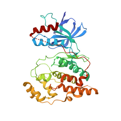
3E7O

Crystal Structure of JNK2


Experimental Data Snapshot

  • Method: X-RAY DIFFRACTION
  • Resolution: 2.14 Å
  • R-Value Free: 0.250 
  • R-Value Work: 0.217 
  • R-Value Observed: 0.219 

Starting Model: experimental
View more details

wwPDB Validation   3D Report Full Report


Ligand Structure Quality Assessment 


This is version 1.4 of the entry. See complete history


Literature

The crystal structure of JNK2 reveals conformational flexibility in the MAP kinase insert and indicates its involvement in the regulation of catalytic activity.

Shaw, D.Wang, S.M.Villasenor, A.G.Tsing, S.Walter, D.Browner, M.F.Barnett, J.Kuglstatter, A.

(2008) J Mol Biol 383: 885-893

  • DOI: https://doi.org/10.1016/j.jmb.2008.08.086
  • Primary Citation of Related Structures:  
    3E7O

  • PubMed Abstract: 

    c-Jun N-terminal kinase (JNK) 2 is a member of the mitogen-activated protein (MAP) kinase group of signaling proteins. MAP kinases share a common sequence insertion called "MAP kinase insert", which, for ERK2, has been shown to interact with regulatory proteins and, for p38alpha, has been proposed to be involved in the regulation of catalytic activity. We have determined the crystal structure of human JNK2 complexed with an indazole inhibitor by applying a high-throughput protein engineering and surface-site mutagenesis approach. A novel conformation of the activation loop is observed, which is not compatible with its phosphorylation by upstream kinases. This activation inhibitory conformation of JNK2 is stabilized by the MAP kinase insert that interacts with the activation loop in an induced-fit manner. We therefore suggest that the MAP kinase insert of JNK2 plays a role in the regulation of JNK2 activation, possibly by interacting with intracellular binding partners.


  • Organizational Affiliation

    Discovery Technologies, Roche Palo Alto, 3431 Hillview Avenue, Palo Alto, CA 94304, USA. david.shaw@roche.com


Macromolecules
Find similar proteins by:  (by identity cutoff)  |  3D Structure
Entity ID: 1
MoleculeChains Sequence LengthOrganismDetailsImage
Mitogen-activated protein kinase 9
A, B
360Homo sapiensMutation(s): 6 
Gene Names: MAPK9JNK2PRKM9
EC: 2.7.11.24
UniProt & NIH Common Fund Data Resources
Find proteins for P45984 (Homo sapiens)
Explore P45984 
Go to UniProtKB:  P45984
PHAROS:  P45984
GTEx:  ENSG00000050748 
Entity Groups  
Sequence Clusters30% Identity50% Identity70% Identity90% Identity95% Identity100% Identity
UniProt GroupP45984
Sequence Annotations
Expand
  • Reference Sequence
Small Molecules
Ligands 1 Unique
IDChains Name / Formula / InChI Key2D Diagram3D Interactions
35F
Query on 35F

Download Ideal Coordinates CCD File 
C [auth A],
D [auth B]
N-{3-[5-(1H-1,2,4-triazol-3-yl)-1H-indazol-3-yl]phenyl}furan-2-carboxamide
C20 H14 N6 O2
LMDMJDCLPIVGQD-UHFFFAOYSA-N
Binding Affinity Annotations 
IDSourceBinding Affinity
35F PDBBind:  3E7O IC50: 120 (nM) from 1 assay(s)
Experimental Data & Validation

Experimental Data

  • Method: X-RAY DIFFRACTION
  • Resolution: 2.14 Å
  • R-Value Free: 0.250 
  • R-Value Work: 0.217 
  • R-Value Observed: 0.219 
  • Space Group: C 1 2 1
Unit Cell:
Length ( Å )Angle ( ˚ )
a = 161.688α = 90
b = 78.784β = 96.43
c = 78.927γ = 90
Software Package:
Software NamePurpose
REFMACrefinement
ADSCdata collection
HKL-2000data reduction
HKL-2000data scaling
PHASERphasing

Structure Validation

View Full Validation Report


Ligand Structure Quality Assessment 


Entry History 

Deposition Data

Revision History  (Full details and data files)

  • Version 1.0: 2008-09-09
    Type: Initial release
  • Version 1.1: 2011-07-13
    Changes: Version format compliance
  • Version 1.2: 2014-04-30
    Changes: Data collection
  • Version 1.3: 2021-10-20
    Changes: Database references, Derived calculations
  • Version 1.4: 2023-08-30
    Changes: Data collection, Refinement description