3EFT | pdb_00003eft

Crystal structure of the complex between Carbonic Anhydrase II and a spin-labeled sulfonamide incorporating TEMPO moiety


Experimental Data Snapshot

  • Method: X-RAY DIFFRACTION
  • Resolution: 1.85 Å
  • R-Value Free: 
    0.240 (Depositor), 0.160 (DCC) 
  • R-Value Work: 
    0.192 (Depositor) 
  • R-Value Observed: 
    0.195 (Depositor) 

Starting Model: experimental
View more details

wwPDB Validation 3D Report Full Report

Validation slider image for 3EFT

Ligand Structure Quality Assessment 


This is version 1.3 of the entry. See complete history

Literature

Dissecting the Inhibition Mechanism of Cytosolic versus Transmembrane Carbonic Anhydrases by ESR

Ciani, L.Cecchi, A.Temperini, C.Supuran, C.T.Ristori, S.

(2009) J Phys Chem B 113: 13998-14005

  • DOI: https://doi.org/10.1021/jp906593c
  • Primary Citation Related Structures: 
    3EFT

  • PubMed Abstract: 

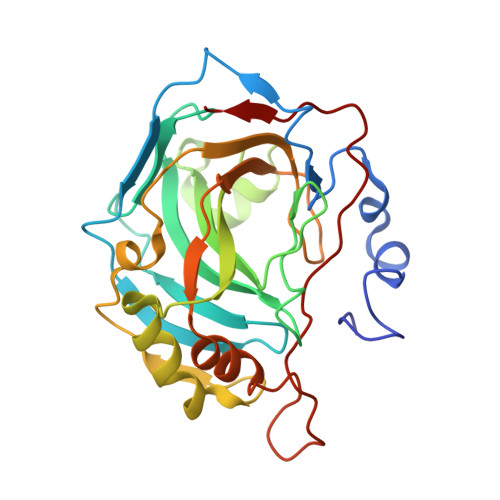
    Spin-labeled sulfonamides incorporating TEMPO moieties showed efficient activity as inhibitors of the metalloenzyme carbonic anhydrase (CA, EC 4.2.1.1) and, in particular, of the physiologically relevant isoenzymes hCA II, hCA IX, and hCA XIV. Here we report a detailed analysis of this class of inhibitors by means of ESR and X-ray crystallography, in comparison with inhibition tests against all mammalian CA isoforms, CA I-XIV. Local dynamics and structure were manifested in the ESR signal through modulation of internal magnetic anisotropies. Analysis and fitting of the ESR spectra of several spin-labeled sulfonamides with isoforms CA II (cytosolic), CA IX (catalytic domain and full length transmembrane, tumor-associated isoform) and CA XIV (transmembrane isozyme) provided information about polarity and dynamics of specific microenvironments sensed by the nitroxyl group within the active site cavity of these isozymes. The comparison of ESR and crystallographic data of hCA II complexed with one of these inhibitors constitutes a useful tool for the understanding of molecular hindrance and ordering within the enzyme active site, and provides theoretical bases to use these inhibitors for imaging purposes of hypoxic tumors overexpressing the transmembrane isozyme CA IX. Combining the sulfonamide zinc-binding group with the TEMPO moiety thus allowed to dissect the selective inhibition mechanism of different cytosolic and transmembrane carbonic anhydrases.


  • Organizational Affiliation
    • Dipartimento di Chimica and CSGI, Universita degli Studi di Firenze, Via della Lastruccia 3, I-50019 Sesto Fiorentino, Firenze, Italy.

Macromolecule Content 

  • Total Structure Weight: 29.98 kDa 
  • Atom Count: 2,323 
  • Modeled Residue Count: 256 
  • Deposited Residue Count: 260 
  • Unique protein chains: 1

Macromolecules

Find similar proteins by:|  3D Structure
Entity ID: 1
MoleculeChains  Sequence LengthOrganismDetailsImage
Carbonic anhydrase 2260N/AMutation(s): 0 
EC: 4.2.1.1 (PDB Primary Data), 4.2.1.69 (UniProt)
UniProt & NIH Common Fund Data Resources
Find proteins for P00918 (Homo sapiens)
Explore P00918 
Go to UniProtKB:  P00918
PHAROS:  P00918
GTEx:  ENSG00000104267 
Entity Groups
Sequence Clusters30% Identity50% Identity70% Identity90% Identity95% Identity100% Identity
UniProt GroupP00918
Sequence Annotations
Expand
Reference Sequence

Small Molecules

Ligands 3 Unique
IDChains Name / Formula / InChI Key2D Diagram3D Interactions
3BS

Query on 3BS


Download:Ideal Coordinates CCD File
D [auth A]3-chloro-4-{[(1-hydroxy-2,2,6,6-tetramethylpiperidin-4-yl)carbamothioyl]amino}benzenesulfonamide
C16 H25 Cl N4 O3 S2
WDEYGBCTTZKGRI-UHFFFAOYSA-N
HG

Query on HG


Download:Ideal Coordinates CCD File
C [auth A]MERCURY (II) ION
Hg
BQPIGGFYSBELGY-UHFFFAOYSA-N
ZN

Query on ZN


Download:Ideal Coordinates CCD File
B [auth A]ZINC ION
Zn
PTFCDOFLOPIGGS-UHFFFAOYSA-N

Experimental Data & Validation

Experimental Data

  • Method: X-RAY DIFFRACTION
  • Resolution: 1.85 Å
  • R-Value Free:  0.240 (Depositor), 0.160 (DCC) 
  • R-Value Work:  0.192 (Depositor) 
  • R-Value Observed: 0.195 (Depositor) 
Space Group: P 1 21 1
Unit Cell:
Length ( Å )Angle ( ˚ )
a = 42.13α = 90
b = 41.5β = 104.3
c = 72.31γ = 90
Software Package:
Software NamePurpose
CrysalisProdata collection
AMoREphasing
REFMACrefinement
CrysalisProdata reduction
CrysalisProdata scaling

Structure Validation

View Full Validation Report


Ligand Structure Quality Assessment 


Entry History 

Deposition Data

Revision History  (Full details and data files)

  • Version 1.0: 2009-09-08
    Type: Initial release
  • Version 1.1: 2011-07-13
    Changes: Version format compliance
  • Version 1.2: 2017-10-25
    Changes: Refinement description
  • Version 1.3: 2023-11-01
    Changes: Data collection, Database references, Derived calculations, Refinement description