3EOB

Crystal structure the Fab fragment of Efalizumab in complex with LFA-1 I domain, Form II


Experimental Data Snapshot

  • Method: X-RAY DIFFRACTION
  • Resolution: 3.60 Å
  • R-Value Free: 0.333 
  • R-Value Work: 0.267 
  • R-Value Observed: 0.269 

wwPDB Validation   3D Report Full Report


This is version 1.1 of the entry. See complete history


Literature

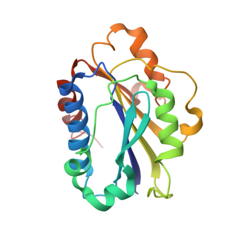
Efalizumab binding to the LFA-1 alphaL I domain blocks ICAM-1 binding via steric hindrance.

Li, S.Wang, H.Peng, B.Zhang, M.Zhang, D.Hou, S.Guo, Y.Ding, J.

(2009) Proc Natl Acad Sci U S A 106: 4349-4354

  • DOI: https://doi.org/10.1073/pnas.0810844106
  • Primary Citation of Related Structures:  
    3EO9, 3EOA, 3EOB

  • PubMed Abstract: 

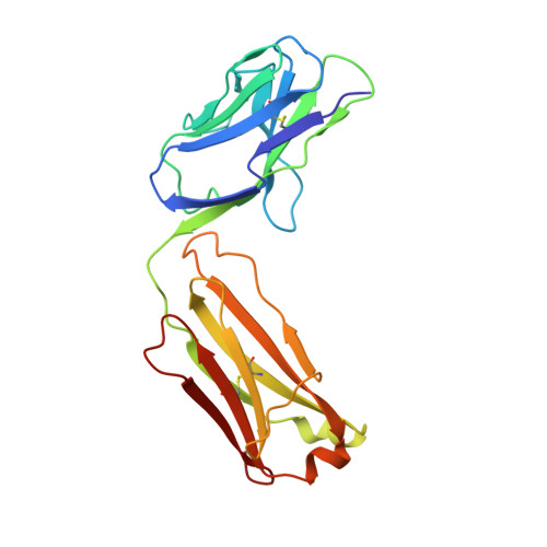
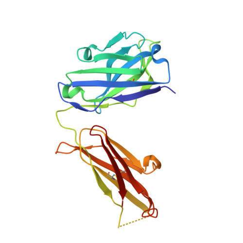
    Lymphocyte function-associated antigen 1 (LFA-1) plays important roles in immune cell adhesion, trafficking, and activation and is a therapeutic target for the treatment of multiple autoimmune diseases. Efalizumab is one of the most efficacious antibody drugs for treating psoriasis, a very common skin disease, through inhibition of the binding of LFA-1 to the ligand intercellular adhesion molecule 1 (ICAM-1). We report here the crystal structures of the Efalizumab Fab alone and in complex with the LFA-1 alpha(L) I domain, which reveal the molecular mechanism of inhibition of LFA-1 by Efalizumab. The Fab binds with an epitope on the inserted (I) domain that is distinct from the ligand-binding site. Efalizumab binding blocks the binding of LFA-1 to ICAM-1 via steric hindrance between its light chain and ICAM-1 domain 2 and thus inhibits the activities of LFA-1. These results have important implications for the development of improved antibodies and new therapeutic strategies for the treatment of autoimmune diseases.


  • Organizational Affiliation

    State Key Laboratory of Molecular Biology and Research Center for Structural Biology, Institute of Biochemistry and Cell Biology, Chinese Academy of Sciences, 320 Yue-Yang Road, Shanghai 200031, China.


Macromolecules
Find similar proteins by:  (by identity cutoff)  |  3D Structure
Entity ID: 1
MoleculeChains Sequence LengthOrganismDetailsImage
Efalizumab Fab fragment, light chainA [auth L],
D [auth A]
214Homo sapiensMutation(s): 0 
Gene Names: IGG1
Entity Groups  
Sequence Clusters30% Identity50% Identity70% Identity90% Identity95% Identity100% Identity
Sequence Annotations
Expand
  • Reference Sequence
Find similar proteins by:  (by identity cutoff)  |  3D Structure
Entity ID: 2
MoleculeChains Sequence LengthOrganismDetailsImage
Efalizumab Fab fragment, heavy chainB [auth H],
E [auth B]
220Homo sapiensMutation(s): 0 
Gene Names: IGG1
Entity Groups  
Sequence Clusters30% Identity50% Identity70% Identity90% Identity95% Identity100% Identity
Sequence Annotations
Expand
  • Reference Sequence
Find similar proteins by:  (by identity cutoff)  |  3D Structure
Entity ID: 3
MoleculeChains Sequence LengthOrganismDetailsImage
Integrin alpha-LC [auth I],
F [auth J]
181Homo sapiensMutation(s): 0 
Gene Names: LFA-1 I domain
UniProt & NIH Common Fund Data Resources
Find proteins for P20701 (Homo sapiens)
Explore P20701 
Go to UniProtKB:  P20701
PHAROS:  P20701
GTEx:  ENSG00000005844 
Entity Groups  
Sequence Clusters30% Identity50% Identity70% Identity90% Identity95% Identity100% Identity
UniProt GroupP20701
Sequence Annotations
Expand
  • Reference Sequence
Experimental Data & Validation

Experimental Data

  • Method: X-RAY DIFFRACTION
  • Resolution: 3.60 Å
  • R-Value Free: 0.333 
  • R-Value Work: 0.267 
  • R-Value Observed: 0.269 
  • Space Group: P 61 2 2
Unit Cell:
Length ( Å )Angle ( ˚ )
a = 111.096α = 90
b = 111.096β = 90
c = 470.726γ = 120
Software Package:
Software NamePurpose
HKL-2000data collection
CNSrefinement
HKL-2000data reduction
HKL-2000data scaling
CNSphasing

Structure Validation

View Full Validation Report


Entry History 

Deposition Data

Revision History  (Full details and data files)

  • Version 1.0: 2009-04-14
    Type: Initial release
  • Version 1.1: 2011-07-13
    Changes: Version format compliance