3EW3

the 1:2 complex between a Nterminal elongated prolactin and the extra cellular domain of the rat prolactin receptor


Experimental Data Snapshot

  • Method: X-RAY DIFFRACTION
  • Resolution: 3.80 Å
  • R-Value Free: 0.324 
  • R-Value Work: 0.252 
  • R-Value Observed: 0.256 

wwPDB Validation   3D Report Full Report


This is version 1.2 of the entry. See complete history


Literature

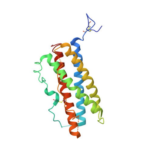
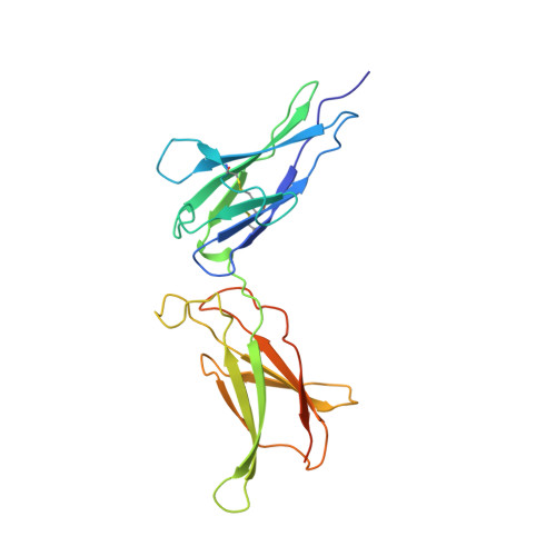
Crystal structure of an affinity-matured prolactin complexed to its dimerized receptor reveals the topology of hormone binding site 2.

Broutin, I.Jomain, J.B.Tallet, E.van Agthoven, J.Raynal, B.Hoos, S.Kragelund, B.B.Kelly, P.A.Ducruix, A.England, P.Goffin, V.

(2010) J Biol Chem 285: 8422-8433

  • DOI: https://doi.org/10.1074/jbc.M109.089128
  • Primary Citation of Related Structures:  
    3EW3

  • PubMed Abstract: 

    We report the first crystal structure of a 1:2 hormone.receptor complex that involves prolactin (PRL) as the ligand, at 3.8-A resolution. Stable ternary complexes were obtained by generating affinity-matured PRL variants harboring an N-terminal tail from ovine placental lactogen, a closely related PRL receptor (PRLR) ligand. This structure allows one to draw up an exhaustive inventory of the residues involved at the PRL.PRLR site 2 interface, consistent with all previously reported site-directed mutagenesis data. We propose, with this description, an interaction model involving three structural components of PRL site 2 ("three-pin plug"): the conserved glycine 129 of helix alpha3, the hydrogen bond network involving surrounding residues (glycine cavity), and the N terminus. The model provides a molecular basis for the properties of the different PRL analogs designed to date, including PRLR antagonists. Finally, comparison of our 1:2 PRL.PRLR(2) structure with those of free PRL and its 1:1 complex indicates that the structure of PRL undergoes significant changes when binding the first, but not the second receptor. This suggests that the second PRLR moiety adapts to the 1:1 complex rather than the opposite. In conclusion, this structure will be a useful guiding tool for further investigations of the molecular mechanisms involved in PRLR dimerization and activation, as well as for the optimization of PRLR antagonists, an emerging class of compounds with high therapeutic potential against breast and prostate cancer.


  • Organizational Affiliation

    Laboratoire de Cristallographie et RMN Biologiques, CNRS, UMR 8015, Université Paris Descartes, 75006 Paris, France.


Macromolecules
Find similar proteins by:  (by identity cutoff)  |  3D Structure
Entity ID: 1
MoleculeChains Sequence LengthOrganismDetailsImage
Prolactin203Homo sapiensMutation(s): 0 
Gene Names: PRL
UniProt & NIH Common Fund Data Resources
Find proteins for P01236 (Homo sapiens)
Explore P01236 
Go to UniProtKB:  P01236
PHAROS:  P01236
GTEx:  ENSG00000172179 
Entity Groups  
Sequence Clusters30% Identity50% Identity70% Identity90% Identity95% Identity100% Identity
UniProt GroupP01236
Sequence Annotations
Expand
  • Reference Sequence
Find similar proteins by:  (by identity cutoff)  |  3D Structure
Entity ID: 2
MoleculeChains Sequence LengthOrganismDetailsImage
Prolactin receptor
B, C
221Rattus norvegicusMutation(s): 0 
Gene Names: Prlr
UniProt
Find proteins for P05710 (Rattus norvegicus)
Explore P05710 
Go to UniProtKB:  P05710
Entity Groups  
Sequence Clusters30% Identity50% Identity70% Identity90% Identity95% Identity100% Identity
UniProt GroupP05710
Sequence Annotations
Expand
  • Reference Sequence
Experimental Data & Validation

Experimental Data

  • Method: X-RAY DIFFRACTION
  • Resolution: 3.80 Å
  • R-Value Free: 0.324 
  • R-Value Work: 0.252 
  • R-Value Observed: 0.256 
  • Space Group: P 43 21 2
Unit Cell:
Length ( Å )Angle ( ˚ )
a = 92.12α = 90
b = 92.12β = 90
c = 215.85γ = 90
Software Package:
Software NamePurpose
MOSFLMdata reduction
SCALAdata scaling
PHASERphasing
CNSrefinement
PDB_EXTRACTdata extraction
ADSCdata collection
PHENIXrefinement

Structure Validation

View Full Validation Report


Entry History 

Deposition Data

Revision History  (Full details and data files)

  • Version 1.0: 2009-11-03
    Type: Initial release
  • Version 1.1: 2011-07-13
    Changes: Version format compliance
  • Version 1.2: 2023-11-01
    Changes: Data collection, Database references, Refinement description