3EZ5

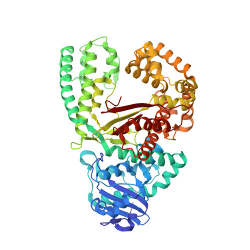
Cocrystal structure of Bacillus fragment DNA polymerase I with duplex DNA , dCTP, and zinc (closed form).


Experimental Data Snapshot

  • Method: X-RAY DIFFRACTION
  • Resolution: 1.90 Å
  • R-Value Free: 0.244 
  • R-Value Work: 0.211 
  • R-Value Observed: 0.212 

Starting Model: experimental
View more details

wwPDB Validation   3D Report Full Report


Ligand Structure Quality Assessment 


This is version 2.2 of the entry. See complete history


Literature

The Mechanism of the Translocation Step in DNA Replication by DNA Polymerase I: A Computer Simulation Analysis.

Golosov, A.A.Warren, J.J.Beese, L.S.Karplus, M.

(2010) Structure 18: 83-93

  • DOI: https://doi.org/10.1016/j.str.2009.10.014
  • Primary Citation of Related Structures:  
    3EYZ, 3EZ5

  • PubMed Abstract: 

    High-fidelity DNA polymerases copy DNA rapidly and accurately by adding correct deoxynucleotide triphosphates to a growing primer strand of DNA. Following nucleotide incorporation, a series of conformational changes translocate the DNA substrate by one base pair step, readying the polymerase for the next round of incorporation. Molecular dynamics simulations indicate that the translocation consists globally of a polymerase fingers-opening transition, followed by the DNA displacement and the insertion of the template base into the preinsertion site. They also show that the pyrophosphate release facilitates the opening transition and that the universally conserved Y714 plays a key role in coupling polymerase opening to DNA translocation. The transition involves several metastable intermediates in one of which the O helix is bent in the vicinity of G711. Completion of the translocation appears to require a gating motion of the O1 helix, perhaps facilitated by the presence of G715. These roles are consistent with the high level of conservation of Y714 and the two glycine residues at these positions. It is likely that a corresponding mechanism is applicable to other polymerases.


  • Organizational Affiliation

    Department of Chemistry and Chemical Biology, Harvard University, 12 Oxford Street, Cambridge, MA 02138, USA.


Macromolecules

Find similar proteins by:  (by identity cutoff)  |  3D Structure
Entity ID: 1
MoleculeChains Sequence LengthOrganismDetailsImage
DNA polymerase IA,
B [auth D]
580N/AMutation(s): 1 
Gene Names: POLApolA
EC: 2.7.7.7
UniProt
Find proteins for P52026 (Geobacillus stearothermophilus)
Explore P52026 
Go to UniProtKB:  P52026
Entity Groups  
Sequence Clusters30% Identity50% Identity70% Identity90% Identity95% Identity100% Identity
UniProt GroupP52026
Sequence Annotations
Expand
  • Reference Sequence

Find similar nucleic acids by:  Sequence   |   3D Structure  

Entity ID: 2
MoleculeChains LengthOrganismImage
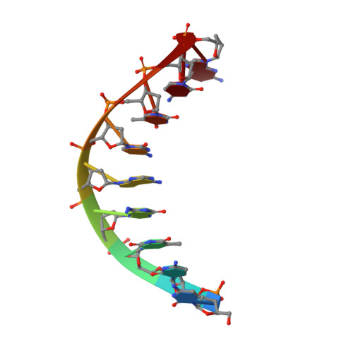
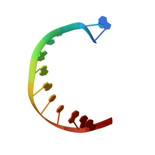
5'-D(*DCP*DCP*DTP*DGP*DAP*DCP*DTP*DCP*DG)-3'C [auth B],
E
9N/A
Sequence Annotations
Expand
  • Reference Sequence

Find similar nucleic acids by:  Sequence   |   3D Structure  

Entity ID: 3
MoleculeChains LengthOrganismImage
5'-D(*DAP*DTP*DTP*DCP*DGP*DAP*DGP*DTP*DCP*DAP*DGP*DG)-3'D [auth C],
F
12N/A
Sequence Annotations
Expand
  • Reference Sequence
Oligosaccharides

Help

Entity ID: 4
MoleculeChains Length2D Diagram Glycosylation3D Interactions
beta-D-fructofuranose-(2-1)-alpha-D-glucopyranose
G, H
2N/A
Glycosylation Resources
GlyTouCan:  G05551OP
GlyCosmos:  G05551OP
Small Molecules
Ligands 3 Unique
IDChains Name / Formula / InChI Key2D Diagram3D Interactions
DAD
Query on DAD

Download Ideal Coordinates CCD File 
M [auth A],
Q [auth D]
2',3'-DIDEOXYADENOSINE-5'-TRIPHOSPHATE
C10 H16 N5 O11 P3
OAKPWEUQDVLTCN-RQJHMYQMSA-N
SO4
Query on SO4

Download Ideal Coordinates CCD File 
I [auth A],
J [auth A],
R [auth D]
SULFATE ION
O4 S
QAOWNCQODCNURD-UHFFFAOYSA-L
ZN
Query on ZN

Download Ideal Coordinates CCD File 
K [auth A],
L [auth A],
N [auth D],
O [auth D],
P [auth D]
ZINC ION
Zn
PTFCDOFLOPIGGS-UHFFFAOYSA-N
Biologically Interesting Molecules (External Reference) 1 Unique
Experimental Data & Validation

Experimental Data

  • Method: X-RAY DIFFRACTION
  • Resolution: 1.90 Å
  • R-Value Free: 0.244 
  • R-Value Work: 0.211 
  • R-Value Observed: 0.212 
  • Space Group: P 21 21 21
Unit Cell:
Length ( Å )Angle ( ˚ )
a = 92.551α = 90
b = 108.572β = 90
c = 149.756γ = 90
Software Package:
Software NamePurpose
XSCALEdata scaling
REFMACrefinement
PDB_EXTRACTdata extraction

Structure Validation

View Full Validation Report


Ligand Structure Quality Assessment 


Entry History 

Deposition Data

Revision History  (Full details and data files)

  • Version 1.0: 2009-11-10
    Type: Initial release
  • Version 1.1: 2011-07-13
    Changes: Version format compliance
  • Version 2.0: 2020-07-29
    Type: Remediation
    Reason: Carbohydrate remediation
    Changes: Advisory, Atomic model, Data collection, Database references, Derived calculations, Non-polymer description, Structure summary
  • Version 2.1: 2021-10-20
    Changes: Database references, Structure summary
  • Version 2.2: 2023-09-06
    Changes: Data collection, Refinement description