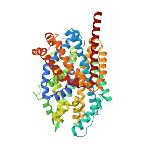
3F4J

Crystal structure of LeuT bound to glycine and sodium


Experimental Data Snapshot

  • Method: X-RAY DIFFRACTION
  • Resolution: 2.15 Å
  • R-Value Free: 0.227 
  • R-Value Work: 0.203 
  • R-Value Observed: 0.205 

Starting Model: experimental
View more details

wwPDB Validation   3D Report Full Report


Ligand Structure Quality Assessment 


This is version 1.4 of the entry. See complete history


Literature

A competitive inhibitor traps LeuT in an open-to-out conformation.

Singh, S.K.Piscitelli, C.L.Yamashita, A.Gouaux, E.

(2008) Science 322: 1655-1661

  • DOI: https://doi.org/10.1126/science.1166777
  • Primary Citation of Related Structures:  
    3F3A, 3F3C, 3F3D, 3F3E, 3F48, 3F4I, 3F4J

  • PubMed Abstract: 

    Secondary transporters are workhorses of cellular membranes, catalyzing the movement of small molecules and ions across the bilayer and coupling substrate passage to ion gradients. However, the conformational changes that accompany substrate transport, the mechanism by which a substrate moves through the transporter, and principles of competitive inhibition remain unclear. We used crystallographic and functional studies on the leucine transporter (LeuT), a model for neurotransmitter sodium symporters, to show that various amino acid substrates induce the same occluded conformational state and that a competitive inhibitor, tryptophan (Trp), traps LeuT in an open-to-out conformation. In the Trp complex, the extracellular gate residues arginine 30 and aspartic acid 404 define a second weak binding site for substrates or inhibitors as they permeate from the extracellular solution to the primary substrate site, which demonstrates how residues that participate in gating also mediate permeation.


  • Organizational Affiliation

    Vollum Institute, Oregon Health and Science University, 3181 Southwest Sam Jackson Park Road, Portland, OR 97239, USA.


Macromolecules
Find similar proteins by:  (by identity cutoff)  |  3D Structure
Entity ID: 1
MoleculeChains Sequence LengthOrganismDetailsImage
Transporter519Aquifex aeolicusMutation(s): 0 
Gene Names: snfaq_2077
Membrane Entity: Yes 
UniProt
Find proteins for O67854 (Aquifex aeolicus (strain VF5))
Explore O67854 
Go to UniProtKB:  O67854
Entity Groups  
Sequence Clusters30% Identity50% Identity70% Identity90% Identity95% Identity100% Identity
UniProt GroupO67854
Sequence Annotations
Expand
  • Reference Sequence
Small Molecules
Ligands 3 Unique
IDChains Name / Formula / InChI Key2D Diagram3D Interactions
BOG
Query on BOG

Download Ideal Coordinates CCD File 
C [auth A],
D [auth A],
E [auth A],
F [auth A]
octyl beta-D-glucopyranoside
C14 H28 O6
HEGSGKPQLMEBJL-RKQHYHRCSA-N
GLY
Query on GLY

Download Ideal Coordinates CCD File 
B [auth A]GLYCINE
C2 H5 N O2
DHMQDGOQFOQNFH-UHFFFAOYSA-N
NA
Query on NA

Download Ideal Coordinates CCD File 
G [auth A],
H [auth A]
SODIUM ION
Na
FKNQFGJONOIPTF-UHFFFAOYSA-N
Experimental Data & Validation

Experimental Data

  • Method: X-RAY DIFFRACTION
  • Resolution: 2.15 Å
  • R-Value Free: 0.227 
  • R-Value Work: 0.203 
  • R-Value Observed: 0.205 
  • Space Group: C 1 2 1
Unit Cell:
Length ( Å )Angle ( ˚ )
a = 90.06α = 90
b = 86.55β = 95.27
c = 81.45γ = 90
Software Package:
Software NamePurpose
PHENIXrefinement
CNSrefinement
ADSCdata collection
HKL-2000data reduction
HKL-2000data scaling
CNSphasing

Structure Validation

View Full Validation Report


Ligand Structure Quality Assessment 


Entry History 

Deposition Data

Revision History  (Full details and data files)

  • Version 1.0: 2008-12-23
    Type: Initial release
  • Version 1.1: 2011-07-13
    Changes: Version format compliance
  • Version 1.2: 2019-07-24
    Changes: Data collection, Refinement description
  • Version 1.3: 2020-07-29
    Type: Remediation
    Reason: Carbohydrate remediation
    Changes: Data collection, Database references, Derived calculations, Structure summary
  • Version 1.4: 2023-09-06
    Changes: Data collection, Database references, Refinement description, Structure summary