3F8E | pdb_00003f8e

Coumarins are a novel class of suicide carbonic anhydrase inhibitors


Experimental Data Snapshot

  • Method: X-RAY DIFFRACTION
  • Resolution: 2.00 Å
  • R-Value Free: 
    0.268 (Depositor), 0.221 (DCC) 
  • R-Value Work: 
    0.199 (Depositor), 0.217 (DCC) 
  • R-Value Observed: 
    0.202 (Depositor) 

Starting Model: experimental
View more details

wwPDB Validation 3D Report Full Report

Validation slider image for 3F8E

Ligand Structure Quality Assessment 


This is version 1.3 of the entry. See complete history

Literature

Non-Zinc Mediated Inhibition of Carbonic Anhydrases: Coumarins Are a New Class of Suicide Inhibitors

Maresca, A.Temperini, C.Vu, H.Pham, N.B.Poulsen, S.-A.Scozzafava, A.Quinn, R.J.Supuran, C.T.

(2009) J Am Chem Soc 131: 3057-3062

  • DOI: https://doi.org/10.1021/ja809683v
  • Primary Citation Related Structures: 
    3F8E

  • PubMed Abstract: 

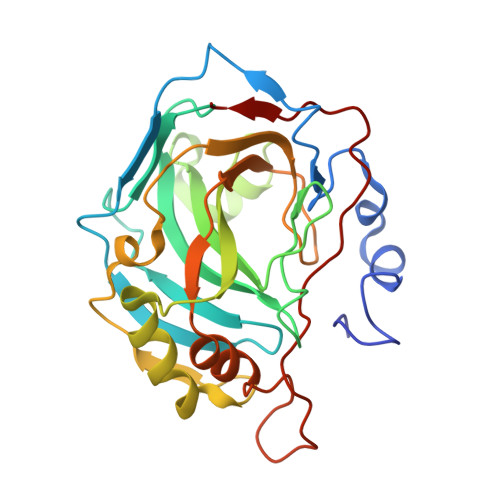
    The X-ray crystal structure of the adduct between the zinc metalloenzyme carbonic anhydrase II (CA, EC 4.2.1.1) with the recently discovered natural product coumarin derivative 6-(1S-hydroxy-3-methylbutyl)-7-methoxy-2H-chromen-2-one showed the coumarin hydrolysis product, a cis-2-hydroxy-cinnamic acid derivative, and not the parent coumarin, bound within the enzyme active site. The bound inhibitor exhibits an extended, two-arm conformation that effectively plugs the entrance to the enzyme active site with no interactions with the catalytically crucial zinc ion. The inhibitor is sandwiched between Phe131, with which it makes an edge-to-face stacking, and Asn67/Glu238sym, with which it makes several polar and hydrogen bonding interactions. This unusual binding mode, with no interactions between the inhibitor molecule and the active site metal ion is previously unobserved for this enzyme class and presents a new opportunity for future drug design campaigns to target a mode of inhibition that differs substantially from classical inhibitors such as the clinically used sulfonamides and sulfamates. Several structurally simple coumarin scaffolds were also shown to inhibit all 13 catalytically active mammalian CA isoforms, with inhibition constants ranging from nanomolar to millimolar. The inhibition is time dependent, with maximum inhibition being observed after 6 h.


  • Organizational Affiliation
    • Università degli Studi di Firenze, Laboratorio di Chimica Bioinorganica, Rm. 188, Via della Lastruccia 3, I-50019 Sesto Fiorentino, Florence, Italy.

Macromolecule Content 

  • Total Structure Weight: 30.16 kDa 
  • Atom Count: 2,281 
  • Modeled Residue Count: 256 
  • Deposited Residue Count: 260 
  • Unique protein chains: 1

Macromolecules

Find similar proteins by:|  3D Structure
Entity ID: 1
MoleculeChains  Sequence LengthOrganismDetailsImage
Carbonic anhydrase 2260Homo sapiensMutation(s): 0 
EC: 4.2.1.1 (PDB Primary Data), 4.2.1.69 (UniProt)
UniProt & NIH Common Fund Data Resources
Find proteins for P00918 (Homo sapiens)
Explore P00918 
Go to UniProtKB:  P00918
PHAROS:  P00918
GTEx:  ENSG00000104267 
Entity Groups
Sequence Clusters30% Identity50% Identity70% Identity90% Identity95% Identity100% Identity
UniProt GroupP00918
Sequence Annotations
Expand
Reference Sequence

Small Molecules

Ligands 4 Unique
IDChains Name / Formula / InChI Key2D Diagram3D Interactions
TE1

Query on TE1


Download:Ideal Coordinates CCD File
E [auth A](2Z)-3-{2-hydroxy-5-[(1S)-1-hydroxy-3-methylbutyl]-4-methoxyphenyl}prop-2-enoic acid
C15 H20 O5
KXEZMFMTEVLUPQ-ZFDPJTLLSA-N
HG

Query on HG


Download:Ideal Coordinates CCD File
C [auth A],
D [auth A]
MERCURY (II) ION
Hg
BQPIGGFYSBELGY-UHFFFAOYSA-N
BEZ

Query on BEZ


Download:Ideal Coordinates CCD File
F [auth A]BENZOIC ACID
C7 H6 O2
WPYMKLBDIGXBTP-UHFFFAOYSA-N
ZN

Query on ZN


Download:Ideal Coordinates CCD File
B [auth A]ZINC ION
Zn
PTFCDOFLOPIGGS-UHFFFAOYSA-N

Experimental Data & Validation

Experimental Data

  • Method: X-RAY DIFFRACTION
  • Resolution: 2.00 Å
  • R-Value Free:  0.268 (Depositor), 0.221 (DCC) 
  • R-Value Work:  0.199 (Depositor), 0.217 (DCC) 
  • R-Value Observed: 0.202 (Depositor) 
Space Group: P 1 21 1
Unit Cell:
Length ( Å )Angle ( ˚ )
a = 42.17α = 90
b = 41.6β = 104.62
c = 72.23γ = 90
Software Package:
Software NamePurpose
CrysalisProdata collection
AMoREphasing
REFMACrefinement
CrysalisProdata reduction
SCALEPACKdata scaling

Structure Validation

View Full Validation Report


Ligand Structure Quality Assessment 


Entry History 

Deposition Data

Revision History  (Full details and data files)

  • Version 1.0: 2009-10-06
    Type: Initial release
  • Version 1.1: 2011-07-13
    Changes: Version format compliance
  • Version 1.2: 2017-11-01
    Changes: Refinement description
  • Version 1.3: 2023-11-01
    Changes: Data collection, Database references, Derived calculations, Refinement description