


Mapping Binding Pocket Volume: Potential Applications towards Ligand Design and Selectivity
Arora, N., Williams, C., Delano, W., Will, D., Kuglstatter, A.To be published.
Experimental Data Snapshot
Starting Model: experimental
View more details
Entity ID: 1 | |||||
|---|---|---|---|---|---|
| Molecule | Chains | Sequence Length | Organism | Details | Image |
| Mitogen-activated protein kinase 14 | 372 | Homo sapiens | Mutation(s): 1 Gene Names: CSBP, CSBP1, CSBP2, CSPB1, MAPK14, MXI2 EC: 2.7.11.24 | 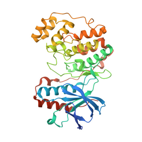 | |
UniProt & NIH Common Fund Data Resources | |||||
PHAROS: Q16539 GTEx: ENSG00000112062 | |||||
Entity Groups | |||||
| Sequence Clusters | 30% Identity50% Identity70% Identity90% Identity95% Identity100% Identity | ||||
| UniProt Group | Q16539 | ||||
Sequence AnnotationsExpand | |||||
Reference Sequence | |||||
| Ligands 1 Unique | |||||
|---|---|---|---|---|---|
| ID | Chains | Name / Formula / InChI Key | 2D Diagram | 3D Interactions | |
| FI4 Download:Ideal Coordinates CCD File | B [auth A] | (2S)-1-{[3-(2-chlorophenyl)-6-(2,4-difluorophenoxy)-1H-pyrazolo[3,4-d]pyrimidin-4-yl]amino}propan-2-ol C20 H16 Cl F2 N5 O2 ULOWCFSJXCABBL-JTQLQIEISA-N |  | ||
| Length ( Å ) | Angle ( ˚ ) |
|---|---|
| a = 45.524 | α = 90 |
| b = 85.828 | β = 90 |
| c = 123.859 | γ = 90 |
| Software Name | Purpose |
|---|---|
| REFMAC | refinement |


