3GIC | pdb_00003gic

Structure of thrombin mutant delta(146-149e) in the free form


Experimental Data Snapshot

  • Method: X-RAY DIFFRACTION
  • Resolution: 1.55 Å
  • R-Value Free: 
    0.220 (Depositor), 0.270 (DCC) 
  • R-Value Work: 
    0.187 (Depositor) 
  • R-Value Observed: 
    0.188 (Depositor) 

Starting Model: experimental
View more details

wwPDB Validation 3D Report Full Report

Validation slider image for 3GIC

Ligand Structure Quality Assessment 


This is version 1.5 of the entry. See complete history

Literature

Stabilization of the E* form turns thrombin into an anticoagulant.

Bah, A.Carrell, C.J.Chen, Z.Gandhi, P.S.Di Cera, E.

(2009) J Biological Chem 284: 20034-20040

  • DOI: https://doi.org/10.1074/jbc.M109.012344
  • Primary Citation Related Structures: 
    3GIC

  • PubMed Abstract: 

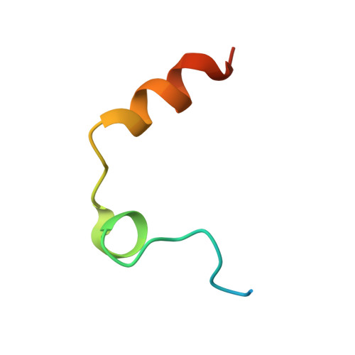
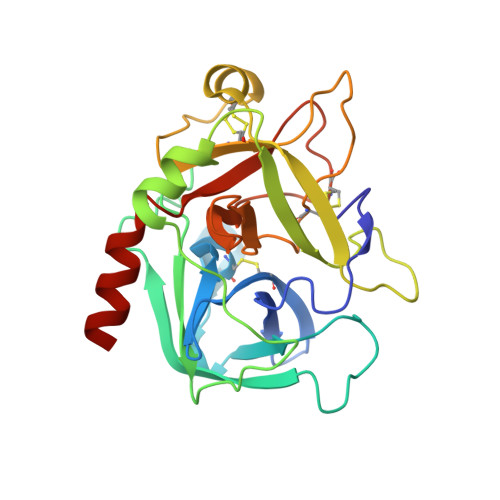
    Previous studies have shown that deletion of nine residues in the autolysis loop of thrombin produces a mutant with an anticoagulant propensity of potential clinical relevance, but the molecular origin of the effect has remained unresolved. The x-ray crystal structure of this mutant solved in the free form at 1.55 A resolution reveals an inactive conformation that is practically identical (root mean square deviation of 0.154 A) to the recently identified E* form. The side chain of Trp(215) collapses into the active site by shifting > 10 A from its position in the active E form, and the oxyanion hole is disrupted by a flip of the Glu(192)-Gly(193) peptide bond. This finding confirms the existence of the inactive form E* in essentially the same incarnation as first identified in the structure of the thrombin mutant D102N. In addition, it demonstrates that the anticoagulant profile often caused by a mutation of the thrombin scaffold finds its likely molecular origin in the stabilization of the inactive E* form that is selectively shifted to the active E form upon thrombomodulin and protein C binding.


  • Organizational Affiliation
    • Department of Biochemistry and Molecular Biophysics, Washington University School of Medicine, St. Louis, Missouri 63110, USA.

Macromolecule Content 

  • Total Structure Weight: 33.11 kDa 
  • Atom Count: 2,577 
  • Modeled Residue Count: 276 
  • Deposited Residue Count: 286 
  • Unique protein chains: 2

Macromolecules

Find similar proteins by:|  3D Structure
Entity ID: 1
MoleculeChains  Sequence LengthOrganismDetailsImage
Thrombin light chain36Homo sapiensMutation(s): 0 
Gene Names: F2
EC: 3.4.21.5
UniProt & NIH Common Fund Data Resources
Find proteins for P00734 (Homo sapiens)
Explore P00734 
Go to UniProtKB:  P00734
PHAROS:  P00734
GTEx:  ENSG00000180210 
Entity Groups
Sequence Clusters30% Identity50% Identity70% Identity90% Identity95% Identity100% Identity
UniProt GroupP00734
Sequence Annotations
Expand
Reference Sequence
Find similar proteins by:|  3D Structure
Entity ID: 2
MoleculeChains  Sequence LengthOrganismDetailsImage
Thrombin heavy chain250Homo sapiensMutation(s): 0 
Gene Names: F2
EC: 3.4.21.5
UniProt & NIH Common Fund Data Resources
Find proteins for P00734 (Homo sapiens)
Explore P00734 
Go to UniProtKB:  P00734
PHAROS:  P00734
GTEx:  ENSG00000180210 
Entity Groups
Sequence Clusters30% Identity50% Identity70% Identity90% Identity95% Identity100% Identity
UniProt GroupP00734
Sequence Annotations
Expand
Reference Sequence

Small Molecules

Ligands 1 Unique
IDChains Name / Formula / InChI Key2D Diagram3D Interactions
NAG

Query on NAG


Download:Ideal Coordinates CCD File
C [auth B]2-acetamido-2-deoxy-beta-D-glucopyranose
C8 H15 N O6
OVRNDRQMDRJTHS-FMDGEEDCSA-N

Experimental Data & Validation

Experimental Data

  • Method: X-RAY DIFFRACTION
  • Resolution: 1.55 Å
  • R-Value Free:  0.220 (Depositor), 0.270 (DCC) 
  • R-Value Work:  0.187 (Depositor) 
  • R-Value Observed: 0.188 (Depositor) 
Space Group: P 43
Unit Cell:
Length ( Å )Angle ( ˚ )
a = 58.229α = 90
b = 58.229β = 90
c = 119.559γ = 90
Software Package:
Software NamePurpose
DENZOdata reduction
SCALEPACKdata scaling
MOLREPphasing
REFMACrefinement
PDB_EXTRACTdata extraction

Structure Validation

View Full Validation Report


Ligand Structure Quality Assessment 


Entry History 

Deposition Data

Revision History  (Full details and data files)

  • Version 1.0: 2009-06-02
    Type: Initial release
  • Version 1.1: 2011-07-13
    Changes: Non-polymer description, Version format compliance
  • Version 1.2: 2017-11-01
    Changes: Refinement description
  • Version 1.3: 2020-07-29
    Type: Remediation
    Reason: Carbohydrate remediation
    Changes: Data collection, Database references, Derived calculations, Structure summary
  • Version 1.4: 2023-09-06
    Changes: Data collection, Database references, Refinement description, Structure summary
  • Version 1.5: 2024-11-27
    Changes: Structure summary