3GXE | pdb_00003gxe

Complex of a Low Affinity Collagen Site with the Fibronectin 8-9FnI Domain Pair


Experimental Data Snapshot

  • Method: X-RAY DIFFRACTION
  • Resolution: 2.60 Å
  • R-Value Free: 
    0.271 (Depositor), 0.270 (DCC) 
  • R-Value Work: 
    0.219 (Depositor), 0.220 (DCC) 
  • R-Value Observed: 
    0.223 (Depositor) 

Starting Model: experimental
View more details

wwPDB Validation   3D Report Full Report


Ligand Structure Quality Assessment 

Created with Raphaël 2.3.0Worse 01 BetterLigand structure goodness of fit to experimental dataBest fitted NAGClick on this verticalbar to view details

This is version 1.7 of the entry. See complete history


Literature

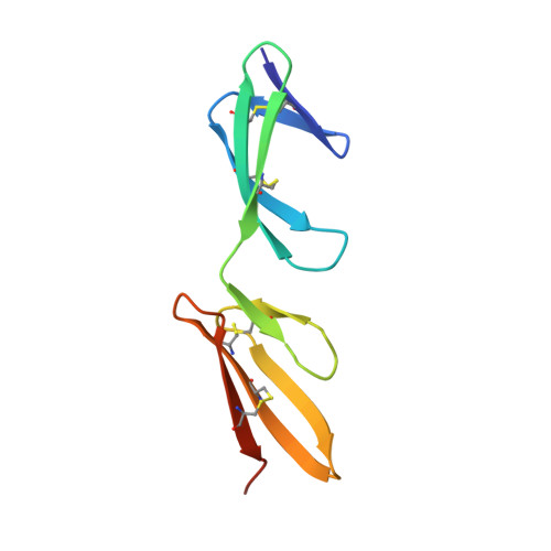
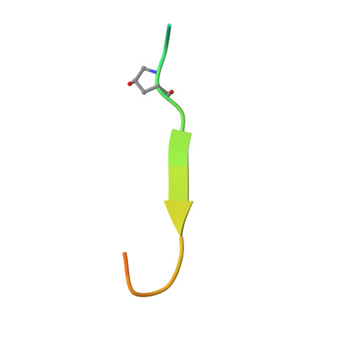
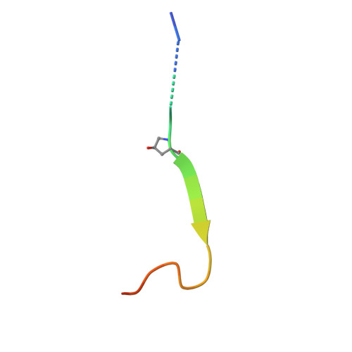
Structural analysis of collagen type I interactions with human fibronectin reveals a cooperative binding mode

Erat, M.C.Sladek, B.Campbell, I.D.Vakonakis, I.

(2013) J Biological Chem 288: 17441-17450

  • DOI: https://doi.org/10.1074/jbc.M113.469841
  • Primary Citation of Related Structures:  
    3GXE

  • PubMed Abstract: 

    Despite its biological importance, the interaction between fibronectin (FN) and collagen, two abundant and crucial tissue components, has not been well characterized on a structural level. Here, we analyzed the four interactions formed between epitopes of collagen type I and the collagen-binding fragment (gelatin-binding domain (GBD)) of human FN using solution NMR, fluorescence, and small angle x-ray scattering methods. Collagen association with FN modules (8-9)FnI occurs through a conserved structural mechanism but exhibits a 400-fold disparity in affinity between collagen sites. This disparity is reduced in the full-length GBD, as (6)FnI(1-2)FnII(7)FnI binds a specific collagen epitope next to the weakest (8-9)FnI-binding site. The cooperative engagement of all GBD modules with collagen results in four broadly equipotent FN-collagen interaction sites. Collagen association stabilizes a distinct monomeric GBD conformation in solution, giving further evidence to the view that FN fragments form well defined functional and structural units.


  • Organizational Affiliation

    Department of Biochemistry, University of Oxford, Oxford OX1 3QU, United Kingdom.


Macromolecules
Find similar proteins by:  (by identity cutoff)  |  3D Structure
Entity ID: 1
MoleculeChains Sequence LengthOrganismDetailsImage
FibronectinA [auth B],
C [auth A]
93Homo sapiensMutation(s): 2 
Gene Names: FN
UniProt & NIH Common Fund Data Resources
Find proteins for P02751 (Homo sapiens)
Explore P02751 
Go to UniProtKB:  P02751
PHAROS:  P02751
GTEx:  ENSG00000115414 
Entity Groups  
Sequence Clusters30% Identity50% Identity70% Identity90% Identity95% Identity100% Identity
UniProt GroupP02751
Glycosylation
Glycosylation Sites: 1Go to GlyGen: P02751-15
Sequence Annotations
Expand
  • Reference Sequence

Find similar proteins by:  Sequence   |   3D Structure  

Entity ID: 2
MoleculeChains Sequence LengthOrganismDetailsImage
Collagen alpha-1(I) chainB [auth F]23N/AMutation(s): 0 
UniProt & NIH Common Fund Data Resources
Find proteins for P02452 (Homo sapiens)
Explore P02452 
Go to UniProtKB:  P02452
PHAROS:  P02452
GTEx:  ENSG00000108821 
Entity Groups  
Sequence Clusters30% Identity50% Identity70% Identity90% Identity95% Identity100% Identity
UniProt GroupP02452
Sequence Annotations
Expand
  • Reference Sequence

Find similar proteins by:  Sequence   |   3D Structure  

Entity ID: 3
MoleculeChains Sequence LengthOrganismDetailsImage
Collagen alpha-1(I) chainD [auth E]23N/AMutation(s): 0 
UniProt & NIH Common Fund Data Resources
Find proteins for P02452 (Homo sapiens)
Explore P02452 
Go to UniProtKB:  P02452
PHAROS:  P02452
GTEx:  ENSG00000108821 
Entity Groups  
Sequence Clusters30% Identity50% Identity70% Identity90% Identity95% Identity100% Identity
UniProt GroupP02452
Sequence Annotations
Expand
  • Reference Sequence
Small Molecules
Modified Residues  2 Unique
IDChains TypeFormula2D DiagramParent
HZP
Query on HZP
B [auth F]L-PEPTIDE LINKINGC5 H9 N O3PRO
HYP
Query on HYP
D [auth E]L-PEPTIDE LINKINGC5 H9 N O3PRO
Experimental Data & Validation

Experimental Data

  • Method: X-RAY DIFFRACTION
  • Resolution: 2.60 Å
  • R-Value Free:  0.271 (Depositor), 0.270 (DCC) 
  • R-Value Work:  0.219 (Depositor), 0.220 (DCC) 
  • R-Value Observed: 0.223 (Depositor) 
Space Group: P 31 2 1
Unit Cell:
Length ( Å )Angle ( ˚ )
a = 56.57α = 90
b = 56.57β = 90
c = 152.661γ = 120
Software Package:
Software NamePurpose
MOSFLMdata reduction
SCALAdata scaling
PHASERphasing
PHENIXrefinement
PDB_EXTRACTdata extraction
ADSCdata collection

Structure Validation

View Full Validation Report


Ligand Structure Quality Assessment 

Created with Raphaël 2.3.0Worse 01 BetterLigand structure goodness of fit to experimental dataBest fitted NAGClick on this verticalbar to view details

Entry History 

Deposition Data

Revision History  (Full details and data files)

  • Version 1.0: 2010-04-07
    Type: Initial release
  • Version 1.1: 2011-07-13
    Changes: Version format compliance
  • Version 1.2: 2013-05-15
    Changes: Database references, Refinement description
  • Version 1.3: 2014-01-29
    Changes: Database references
  • Version 1.4: 2020-07-29
    Type: Remediation
    Reason: Carbohydrate remediation
    Changes: Data collection, Database references, Derived calculations, Structure summary
  • Version 1.5: 2021-11-10
    Changes: Database references, Structure summary
  • Version 1.6: 2023-11-01
    Changes: Data collection, Refinement description
  • Version 1.7: 2023-11-22
    Changes: Data collection