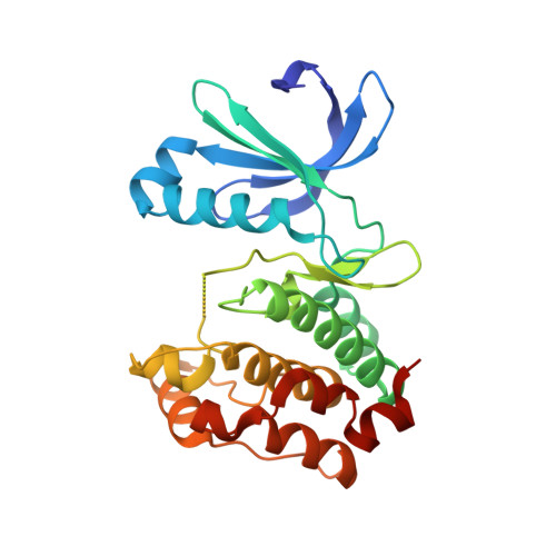
3H0Y

Aurora A in complex with a bisanilinopyrimidine


Experimental Data Snapshot

  • Method: X-RAY DIFFRACTION
  • Resolution: 2.50 Å
  • R-Value Free: 0.285 
  • R-Value Work: 0.208 
  • R-Value Observed: 0.216 

wwPDB Validation   3D Report Full Report


Ligand Structure Quality Assessment 


This is version 1.3 of the entry. See complete history


Literature

A class of 2,4-bisanilinopyrimidine Aurora A inhibitors with unusually high selectivity against Aurora B.

Aliagas-Martin, I.Burdick, D.Corson, L.Dotson, J.Drummond, J.Fields, C.Huang, O.W.Hunsaker, T.Kleinheinz, T.Krueger, E.Liang, J.Moffat, J.Phillips, G.Pulk, R.Rawson, T.E.Ultsch, M.Walker, L.Wiesmann, C.Zhang, B.Zhu, B.Y.Cochran, A.G.

(2009) J Med Chem 52: 3300-3307

  • DOI: https://doi.org/10.1021/jm9000314
  • Primary Citation of Related Structures:  
    3H0Y, 3H0Z, 3H10

  • PubMed Abstract: 

    The two major Aurora kinases carry out critical functions at distinct mitotic stages. Selective inhibitors of these kinases, as well as pan-Aurora inhibitors, show antitumor efficacy and are now under clinical investigation. However, the ATP-binding sites of Aurora A and Aurora B are virtually identical, and the structural basis for selective inhibition has therefore not been clear. We report here a class of bisanilinopyrimidine Aurora A inhibitors with excellent selectivity for Aurora A over Aurora B, both in enzymatic assays and in cellular phenotypic assays. Crystal structures of two of the inhibitors in complex with Aurora A implicate a single amino acid difference in Aurora B as responsible for poor inhibitory activity against this enzyme. Mutation of this residue in Aurora B (E161T) or Aurora A (T217E) is sufficient to swap the inhibition profile, suggesting that this difference might be exploited more generally to achieve high selectivity for Aurora A.


  • Organizational Affiliation

    Department of Small Molecule Drug Discovery, Genentech, Inc., 1 DNA Way, South San Francisco, California 94080, USA.


Macromolecules
Find similar proteins by:  (by identity cutoff)  |  3D Structure
Entity ID: 1
MoleculeChains Sequence LengthOrganismDetailsImage
Serine/threonine-protein kinase 6268Homo sapiensMutation(s): 3 
Gene Names: AURKAAIKARK1AURABTAKSTK15STK6
EC: 2.7.11.1
UniProt & NIH Common Fund Data Resources
Find proteins for O14965 (Homo sapiens)
Explore O14965 
Go to UniProtKB:  O14965
PHAROS:  O14965
GTEx:  ENSG00000087586 
Entity Groups  
Sequence Clusters30% Identity50% Identity70% Identity90% Identity95% Identity100% Identity
UniProt GroupO14965
Sequence Annotations
Expand
  • Reference Sequence
Experimental Data & Validation

Experimental Data

  • Method: X-RAY DIFFRACTION
  • Resolution: 2.50 Å
  • R-Value Free: 0.285 
  • R-Value Work: 0.208 
  • R-Value Observed: 0.216 
  • Space Group: P 61 2 2
Unit Cell:
Length ( Å )Angle ( ˚ )
a = 81.377α = 90
b = 81.377β = 90
c = 170.892γ = 120
Software Package:
Software NamePurpose
HKL-2000data collection
AMoREphasing
REFMACrefinement
DENZOdata reduction
SCALEPACKdata scaling

Structure Validation

View Full Validation Report


Ligand Structure Quality Assessment 


Entry History 

Deposition Data

Revision History  (Full details and data files)

  • Version 1.0: 2009-07-07
    Type: Initial release
  • Version 1.1: 2011-07-13
    Changes: Advisory, Refinement description, Version format compliance
  • Version 1.2: 2021-10-13
    Changes: Database references, Derived calculations
  • Version 1.3: 2024-02-21
    Changes: Data collection