RCSB PDB - 3H3B: Crystal structure of the single-chain Fv (scFv) fragment of an anti-ErbB2 antibody chA21 in complex with residues 1-192 of ErbB2 extracellular domain

 3H3B

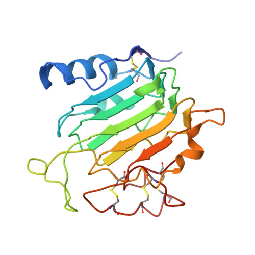
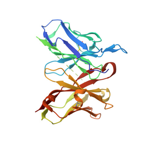
Crystal structure of the single-chain Fv (scFv) fragment of an anti-ErbB2 antibody chA21 in complex with residues 1-192 of ErbB2 extracellular domain


Experimental Data Snapshot

  • Method: X-RAY DIFFRACTION
  • Resolution: 2.45 Å
  • R-Value Free: 0.255 
  • R-Value Work: 0.201 
  • R-Value Observed: 0.203 

Starting Models: experimental
View more details

wwPDB Validation   3D Report Full Report


This is version 1.4 of the entry. See complete history


Literature

Structural Insights into the Down-regulation of Overexpressed p185her2/neu Protein of Transformed Cells by the Antibody chA21.

Zhou, H.Zha, Z.Liu, Y.Zhang, H.Zhu, J.Hu, S.Shen, G.Cheng, L.Niu, L.Greene, M.I.Teng, M.Liu, J.

(2011) J Biol Chem 286: 31676-31683

  • DOI: https://doi.org/10.1074/jbc.M111.235184
  • Primary Citation of Related Structures:  
    3H3B

  • PubMed Abstract: 

    p185(her2/neu) belongs to the ErbB receptor tyrosine kinase family, which has been associated with human breast, ovarian, and lung cancers. Targeted therapies employing ectodomain-specific p185(her2/neu) monoclonal antibodies (mAbs) have demonstrated clinical efficacy for breast cancer. Our previous studies have shown that p185(her2/neu) mAbs are able to disable the kinase activity of homomeric and heteromeric kinase complexes and induce the conversion of the malignant to normal phenotype. We previously developed a chimeric antibody chA21 that specifically inhibits the growth of p185(her2/neu)-overexpressing cancer cells in vitro and in vivo. Herein, we report the crystal structure of the single-chain Fv of chA21 in complex with an N-terminal fragment of p185(her2/neu), which reveals that chA21 binds a region opposite to the dimerization interface, indicating that chA21 does not directly disrupt the dimerization. In contrast, the bivalent chA21 leads to internalization and down-regulation of p185(her2/neu). We propose a structure-based model in which chA21 cross-links two p185(her2/neu) molecules on separate homo- or heterodimers to form a large oligomer in the cell membrane. This model reveals a mechanism for mAbs to drive the receptors into the internalization/degradation path from the inactive hypophosphorylated tetramers formed dynamically by active dimers during a "physiologic process."


  • Organizational Affiliation

    School of Life Sciences, Hefei National Laboratory for Physical Sciences at Microscale, Chinese Academy of Sciences, University of Science and Technology of China, 96 Jinzhai Road, Hefei, Anhui 230027, China.


Macromolecules
Find similar proteins by:  (by identity cutoff)  |  3D Structure
Entity ID: 1
MoleculeChains Sequence LengthOrganismDetailsImage
Receptor tyrosine-protein kinase erbB-2
A, B
194Homo sapiensMutation(s): 0 
Gene Names: ERBB2
EC: 2.7.10.1
UniProt & NIH Common Fund Data Resources
Find proteins for P04626 (Homo sapiens)
Explore P04626 
Go to UniProtKB:  P04626
PHAROS:  P04626
GTEx:  ENSG00000141736 
Entity Groups  
Sequence Clusters30% Identity50% Identity70% Identity90% Identity95% Identity100% Identity
UniProt GroupP04626
Sequence Annotations
Expand
  • Reference Sequence
Find similar proteins by:  (by identity cutoff)  |  3D Structure
Entity ID: 2
MoleculeChains Sequence LengthOrganismDetailsImage
anti-ErbB2 antibody chA21
C, D
259Mus musculusMutation(s): 0 
Gene Names: scFv of murine antibody A21
Entity Groups  
Sequence Clusters30% Identity50% Identity70% Identity90% Identity95% Identity100% Identity
Sequence Annotations
Expand
  • Reference Sequence
Experimental Data & Validation

Experimental Data

  • Method: X-RAY DIFFRACTION
  • Resolution: 2.45 Å
  • R-Value Free: 0.255 
  • R-Value Work: 0.201 
  • R-Value Observed: 0.203 
  • Space Group: P 21 21 21
Unit Cell:
Length ( Å )Angle ( ˚ )
a = 82.177α = 90
b = 87.2β = 90
c = 108.52γ = 90
Software Package:
Software NamePurpose
MAR345dtbdata collection
PHASERphasing
REFMACrefinement
MOSFLMdata reduction
SCALAdata scaling

Structure Validation

View Full Validation Report


Entry History 

Deposition Data

Revision History  (Full details and data files)

  • Version 1.0: 2010-04-28
    Type: Initial release
  • Version 1.1: 2011-07-13
    Changes: Advisory, Version format compliance
  • Version 1.2: 2011-09-28
    Changes: Database references
  • Version 1.3: 2023-11-01
    Changes: Data collection, Database references, Refinement description
  • Version 1.4: 2024-10-30
    Changes: Structure summary