3HF8 | pdb_00003hf8

Crystal structure of human tryoptophan hydroxylase type 1 with bound LP-533401 and Fe


Experimental Data Snapshot

  • Method: X-RAY DIFFRACTION
  • Resolution: 1.85 Å
  • R-Value Free: 
    0.241 (Depositor) 
  • R-Value Work: 
    0.197 (Depositor), 0.198 (DCC) 
  • R-Value Observed: 
    0.199 (Depositor) 

wwPDB Validation 3D Report Full Report

Validation slider image for 3HF8

Ligand Structure Quality Assessment 


This is version 1.3 of the entry. See complete history

Literature

Mechanism of Inhibition of Novel Tryptophan Hydroxylase Inhibitors Revealed by Co-crystal Structures and Kinetic Analysis.

Cianchetta, G.Stouch, T.Yu, W.Shi, Z.C.Tari, L.W.Swanson, R.V.Hunter, M.J.Hoffman, I.D.Liu, Q.

(2010) Curr Chem Genomics 4: 19-26

  • DOI: https://doi.org/10.2174/1875397301004010019
  • Primary Citation Related Structures: 
    3HF8, 3HFB

  • PubMed Abstract: 

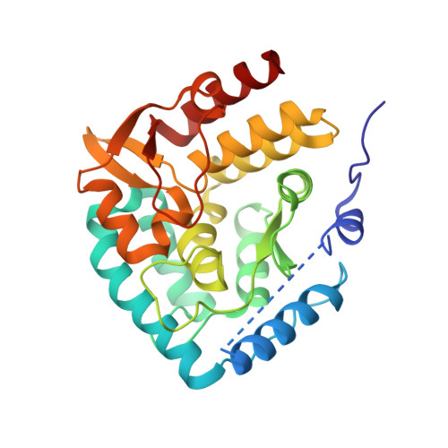
    Trytophan Hydroxylase Type I (TPH1), most abundantly expressed in the gastrointestinal tract, initiates the synthesis of serotonin by catalyzing hydroxylation of tryptophan in the presence of biopterin and oxygen. We have previously described three series of novel, periphery-specific TPH1 inhibitors that selectively deplete serotonin in the gastrointestinal tract. We have now determined co-crystal structures of TPH1 with three of these inhibitors at high resolution. Analysis of the structural data showed that each of the three inhibitors fills the tryptophan binding pocket of TPH1 without reaching into the binding site of the cofactor pterin, and induces major conformational changes of the enzyme. The enzyme-inhibitor complexes assume a compact conformation that is similar to the one in tryptophan complex. Kinetic analysis showed that all three inhibitors are competitive versus the substrate tryptophan, consistent with the structural data that the compounds occupy the tryptophan binding site. On the other hand, all three inhibitors appear to be uncompetitive versus the cofactor 6-methyltetrahydropterin, which is not only consistent with the structural data but also indicate that the hydroxylation reaction follows an ordered binding mechanism in which a productive complex is formed only if tryptophan binds only after pterin, similar to the kinetic mechanisms of tyrosine and phenylalanine hydroxylase.


  • Organizational Affiliation
    • Department of Medicinal Chemistry, Lexicon Pharmaceuticals, Inc., 350 Carter Rd., Princeton, New Jersey, USA.

Macromolecule Content 

  • Total Structure Weight: 33.87 kDa 
  • Atom Count: 2,696 
  • Modeled Residue Count: 274 
  • Deposited Residue Count: 290 
  • Unique protein chains: 1

Macromolecules

Find similar proteins by:|  3D Structure
Entity ID: 1
MoleculeChains  Sequence LengthOrganismDetailsImage
Tryptophan 5-hydroxylase 1290Homo sapiensMutation(s): 0 
EC: 1.14.16.4
UniProt & NIH Common Fund Data Resources
Find proteins for P17752 (Homo sapiens)
Explore P17752 
Go to UniProtKB:  P17752
PHAROS:  P17752
GTEx:  ENSG00000129167 
Entity Groups
Sequence Clusters30% Identity50% Identity70% Identity90% Identity95% Identity100% Identity
UniProt GroupP17752
Sequence Annotations
Expand
Reference Sequence

Small Molecules

Ligands 2 Unique
IDChains Name / Formula / InChI Key2D Diagram3D Interactions
ML0

Query on ML0


Download:Ideal Coordinates CCD File
C [auth A]4-{2-amino-6-[(1R)-2,2,2-trifluoro-1-(3'-fluorobiphenyl-4-yl)ethoxy]pyrimidin-4-yl}-L-phenylalanine
C27 H22 F4 N4 O3
JZWUKILTKYJLCN-XUZZJYLKSA-N
FE

Query on FE


Download:Ideal Coordinates CCD File
B [auth A]FE (III) ION
Fe
VTLYFUHAOXGGBS-UHFFFAOYSA-N

Experimental Data & Validation

Experimental Data

  • Method: X-RAY DIFFRACTION
  • Resolution: 1.85 Å
  • R-Value Free:  0.241 (Depositor) 
  • R-Value Work:  0.197 (Depositor), 0.198 (DCC) 
  • R-Value Observed: 0.199 (Depositor) 
Space Group: P 1 21 1
Unit Cell:
Length ( Å )Angle ( ˚ )
a = 47.278α = 90
b = 58.238β = 97.15
c = 56.554γ = 90
Software Package:
Software NamePurpose
REFMACrefinement

Structure Validation

View Full Validation Report


Ligand Structure Quality Assessment 


Entry History 

Deposition Data

Revision History  (Full details and data files)

  • Version 1.0: 2010-04-21
    Type: Initial release
  • Version 1.1: 2011-07-13
    Changes: Version format compliance
  • Version 1.2: 2012-03-28
    Changes: Database references
  • Version 1.3: 2024-02-21
    Changes: Data collection, Database references, Derived calculations