3HI6 | pdb_00003hi6

Crystal structure of intermediate affinity I domain of integrin LFA-1 with the Fab fragment of its antibody AL-57


Experimental Data Snapshot

  • Method: X-RAY DIFFRACTION
  • Resolution: 2.30 Å
  • R-Value Free: 
    0.226 (Depositor), 0.259 (DCC) 
  • R-Value Work: 
    0.170 (Depositor), 0.224 (DCC) 
  • R-Value Observed: 
    0.173 (Depositor) 

wwPDB Validation 3D Report Full Report

Validation slider image for 3HI6

This is version 1.4 of the entry. See complete history

Literature

Structural basis of activation-dependent binding of ligand-mimetic antibody AL-57 to integrin LFA-1.

Zhang, H.Liu, J.H.Yang, W.Springer, T.Shimaoka, M.Wang, J.H.

(2009) Proc Natl Acad Sci U S A 106: 18345-18350

  • DOI: https://doi.org/10.1073/pnas.0909301106
  • Primary Citation Related Structures: 
    3HI5, 3HI6

  • PubMed Abstract: 

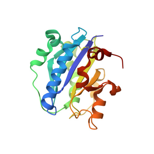
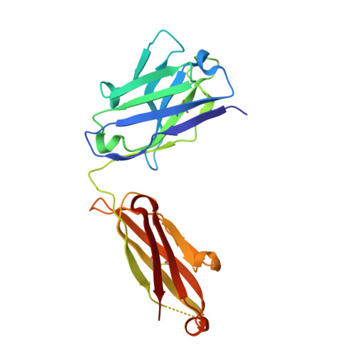
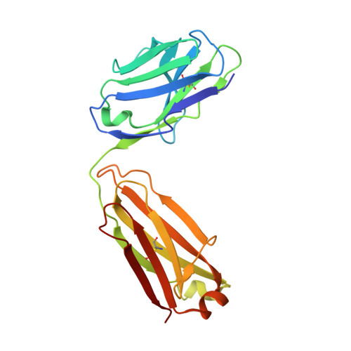
    The activity of integrin LFA-1 (alpha(L)beta(2)) to its ligand ICAM-1 is regulated through the conformational changes of its ligand-binding domain, the I domain of alpha(L) chain, from an inactive, low-affinity closed form (LA), to an intermediate-affinity form (IA), and then finally, to a high-affinity open form (HA). A ligand-mimetic human monoclonal antibody AL-57 (activated LFA-1 clone 57) was identified by phage display to specifically recognize the affinity-upregulated I domain. Here, we describe the crystal structures of the Fab fragment of AL-57 in complex with IA, as well as in its unligated form. We discuss the structural features conferring AL-57's strong selectivity for the high affinity, open conformation of the I domain. The AL-57-binding site overlaps the ICAM-1 binding site on the I domain. Furthermore, an antibody Asp mimics an ICAM Glu by forming a coordination to the metal-ion dependent adhesion site (MIDAS). The structure also reveals better shape complementarity and a more hydrophobic interacting interface in AL-57 binding than in ICAM-1 binding. The results explain AL-57's antagonistic mimicry of LFA-1's natural ligands, the ICAM molecules.


  • Organizational Affiliation
    • Department of Pediatrics, Dana-Farber Cancer Institute, Harvard Medical School, Boston, MA 02115, USA.

Macromolecule Content 

  • Total Structure Weight: 135.36 kDa 
  • Atom Count: 10,097 
  • Modeled Residue Count: 1,209 
  • Deposited Residue Count: 1,224 
  • Unique protein chains: 3

Macromolecules

Find similar proteins by:|  3D Structure
Entity ID: 1
MoleculeChains  Sequence LengthOrganismDetailsImage
Integrin alpha-L
A, B
180Homo sapiensMutation(s): 3 
Gene Names: CD11AITGALLFA-1
UniProt & NIH Common Fund Data Resources
Find proteins for P20701 (Homo sapiens)
Explore P20701 
Go to UniProtKB:  P20701
PHAROS:  P20701
GTEx:  ENSG00000005844 
Entity Groups
Sequence Clusters30% Identity50% Identity70% Identity90% Identity95% Identity100% Identity
UniProt GroupP20701
Sequence Annotations
Expand
Reference Sequence
Find similar proteins by:|  3D Structure
Entity ID: 2
MoleculeChains  Sequence LengthOrganismDetailsImage
Heavy chain of Fab fragment of AL-57 against alpha L I domainC [auth H],
E [auth X]
220Homo sapiensMutation(s): 0 
Find similar proteins by:|  3D Structure
Entity ID: 3
MoleculeChains  Sequence LengthOrganismDetailsImage
light chain of Fab fragment of AL-57 against alpha L I domainD [auth L],
F [auth Y]
212Homo sapiensMutation(s): 0 

Small Molecules

Ligands 2 Unique
IDChains Name / Formula / InChI Key2D Diagram3D Interactions
SO4

Query on SO4


Download:Ideal Coordinates CCD File
I [auth B]
J [auth H]
K [auth H]
L
M [auth X]
I [auth B],
J [auth H],
K [auth H],
L,
M [auth X],
N [auth Y]
SULFATE ION
O4 S
QAOWNCQODCNURD-UHFFFAOYSA-L
MN

Query on MN


Download:Ideal Coordinates CCD File
G [auth A],
H [auth B]
MANGANESE (II) ION
Mn
WAEMQWOKJMHJLA-UHFFFAOYSA-N

Experimental Data & Validation

Experimental Data

  • Method: X-RAY DIFFRACTION
  • Resolution: 2.30 Å
  • R-Value Free:  0.226 (Depositor), 0.259 (DCC) 
  • R-Value Work:  0.170 (Depositor), 0.224 (DCC) 
  • R-Value Observed: 0.173 (Depositor) 
Space Group: P 65
Unit Cell:
Length ( Å )Angle ( ˚ )
a = 133.78α = 90
b = 133.78β = 90
c = 161.05γ = 120
Software Package:
Software NamePurpose
DENZOdata reduction
SCALEPACKdata scaling
MOLREPphasing
REFMACrefinement
PDB_EXTRACTdata extraction
ADSCdata collection

Structure Validation

View Full Validation Report


Entry History 

Deposition Data

Revision History  (Full details and data files)

  • Version 1.0: 2009-09-22
    Type: Initial release
  • Version 1.1: 2011-07-13
    Changes: Version format compliance
  • Version 1.2: 2017-11-01
    Changes: Refinement description
  • Version 1.3: 2021-10-13
    Changes: Database references, Derived calculations
  • Version 1.4: 2024-10-16
    Changes: Data collection, Structure summary