3HON

Crystal Structure of Human Collagen XVIII Trimerization Domain (cubic form)


Experimental Data Snapshot

  • Method: X-RAY DIFFRACTION
  • Resolution: 3.00 Å
  • R-Value Free: 0.288 
  • R-Value Work: 0.279 
  • R-Value Observed: 0.279 

wwPDB Validation   3D Report Full Report


This is version 1.4 of the entry. See complete history


Literature

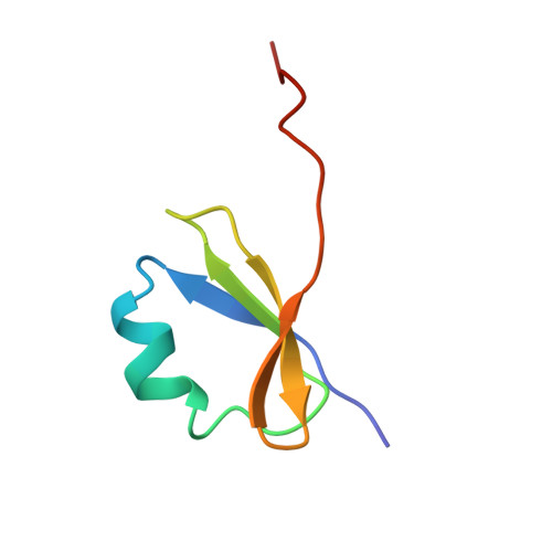
Crystal structure of human collagen XVIII trimerization domain: A novel collagen trimerization Fold.

Boudko, S.P.Sasaki, T.Engel, J.Lerch, T.F.Nix, J.Chapman, M.S.Bachinger, H.P.

(2009) J Mol Biol 392: 787-802

  • DOI: https://doi.org/10.1016/j.jmb.2009.07.057
  • Primary Citation of Related Structures:  
    3HON, 3HSH

  • PubMed Abstract: 

    Collagens contain a unique triple-helical structure with a repeating sequence -G-X-Y-, where proline and hydroxyproline are major constituents in X and Y positions, respectively. Folding of the collagen triple helix requires trimerization domains. Once trimerized, collagen chains are correctly aligned and the folding of the triple helix proceeds in a zipper-like fashion. Here we report the isolation, characterization, and crystal structure of the trimerization domain of human type XVIII collagen, a member of the multiplexin family. This domain differs from all other known trimerization domains in other collagens and exhibits a high trimerization potential at picomolar concentrations. Strong chain association and high specificity of binding are needed for multiplexins, which are present at very low levels.


  • Organizational Affiliation

    Research Department of Shriners Hospital for Children, Portland, OR 97239, USA.


Macromolecules
Find similar proteins by:  (by identity cutoff)  |  3D Structure
Entity ID: 1
MoleculeChains Sequence LengthOrganismDetailsImage
Collagen alpha-1(XVIII) chain56Homo sapiensMutation(s): 1 
Gene Names: COL18A1
UniProt & NIH Common Fund Data Resources
Find proteins for P39060 (Homo sapiens)
Explore P39060 
Go to UniProtKB:  P39060
PHAROS:  P39060
GTEx:  ENSG00000182871 
Entity Groups  
Sequence Clusters30% Identity50% Identity70% Identity90% Identity95% Identity100% Identity
UniProt GroupP39060
Sequence Annotations
Expand
  • Reference Sequence
Experimental Data & Validation

Experimental Data

  • Method: X-RAY DIFFRACTION
  • Resolution: 3.00 Å
  • R-Value Free: 0.288 
  • R-Value Work: 0.279 
  • R-Value Observed: 0.279 
  • Space Group: I 41 3 2
Unit Cell:
Length ( Å )Angle ( ˚ )
a = 94.876α = 90
b = 94.876β = 90
c = 94.876γ = 90
Software Package:
Software NamePurpose
JBluIce-EPICSdata collection
Blu-Icedata collection
PHENIXmodel building
CNSrefinement
MOSFLMdata reduction
SCALAdata scaling
PHENIXphasing

Structure Validation

View Full Validation Report


Entry History 

Deposition Data

Revision History  (Full details and data files)

  • Version 1.0: 2009-08-11
    Type: Initial release
  • Version 1.1: 2011-07-13
    Changes: Version format compliance
  • Version 1.2: 2017-11-01
    Changes: Refinement description
  • Version 1.3: 2018-03-14
    Changes: Database references
  • Version 1.4: 2024-02-21
    Changes: Data collection, Database references