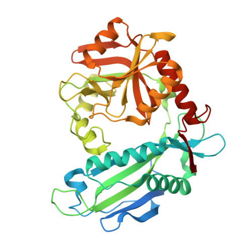
The 1.9 A structure of the branched-chain amino-acid transaminase (IlvE) from Mycobacterium tuberculosis.
Tremblay, L.W., Blanchard, J.S.(2009) Acta Crystallogr Sect F Struct Biol Cryst Commun 65: 1071-1077
- PubMed: 19923721
- DOI: https://doi.org/10.1107/S1744309109036690
- Primary Citation of Related Structures:
3HT5 - PubMed Abstract:
Unlike mammals, bacteria encode enzymes that synthesize branched-chain amino acids. The pyridoxal 50-phosphate-dependent transaminase performs the final biosynthetic step in these pathways, converting keto acid precursors into -amino acids. The branched-chain amino-acid transaminase from Mycobacterium tuberculosis (MtIlvE) has been crystallized and its structure has been solved at 1.9 angstrom resolution. The MtIlvE monomer is composed of two domains that interact to form the active site. The biologically active form of IlvE is a homodimer in which each monomer contributes a substrate-specificity loop to the partner molecule. Additional substrate selectivity may be imparted by a conserved N-terminal Phe30 residue, which has previously been observed to shield the active site in the type IV fold homodimer. The active site of MtIlvE contains density corresponding to bound PMP, which is likely to be a consequence of the presence of tryptone in the crystallization medium. Additionally, two cysteine residues are positioned at the dimer interface for disulfide-bond formation under oxidative conditions. It is unknown whether they are involved in any regulatory activities analogous to those of the human mitochondrial branched-chain amino-acid transaminase.
- Department of Biochemistry, Albert Einstein College of Medicine, Bronx, NY 10461, USA.
Organizational Affiliation:


