


Crystal structure of human PDE4a with 4-(3-butoxy-4-methoxyphenyl)methyl-2-imidazolidone
Cheng, R.K.Y., Crawley, L., Barker, J., Wood, M., Felicetti, B., Whittaker, M.To be published.
Experimental Data Snapshot
Starting Model: experimental
View more details
Entity ID: 1 | |||||
|---|---|---|---|---|---|
| Molecule | Chains | Sequence Length | Organism | Details | Image |
| cAMP-specific 3',5'-cyclic phosphodiesterase 4A | 354 | Homo sapiens | Mutation(s): 0 Gene Names: PDE4A, DPDE2 EC: 3.1.4.17 (PDB Primary Data), 3.1.4.53 (UniProt) | 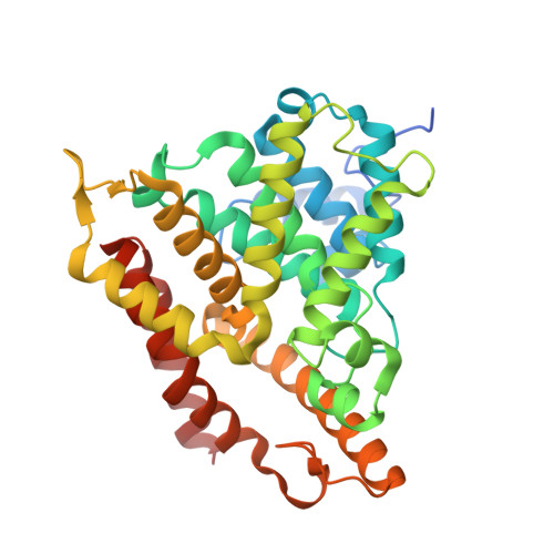 | |
UniProt & NIH Common Fund Data Resources | |||||
PHAROS: P27815 GTEx: ENSG00000065989 | |||||
Entity Groups | |||||
| Sequence Clusters | 30% Identity50% Identity70% Identity90% Identity95% Identity100% Identity | ||||
| UniProt Group | P27815 | ||||
Sequence AnnotationsExpand | |||||
Reference Sequence | |||||
| Ligands 5 Unique | |||||
|---|---|---|---|---|---|
| ID | Chains | Name / Formula / InChI Key | 2D Diagram | 3D Interactions | |
| 0MO Download:Ideal Coordinates CCD File | E [auth A], J [auth B] | (4R)-4-(3-butoxy-4-methoxybenzyl)imidazolidin-2-one C15 H22 N2 O3 PDMUULPVBYQBBK-GFCCVEGCSA-N |  | ||
| GOL Download:Ideal Coordinates CCD File | F [auth A], K [auth B], L [auth B] | GLYCEROL C3 H8 O3 PEDCQBHIVMGVHV-UHFFFAOYSA-N |  | ||
| ZN Download:Ideal Coordinates CCD File | C [auth A], H [auth B] | ZINC ION Zn PTFCDOFLOPIGGS-UHFFFAOYSA-N |  | ||
| CO Download:Ideal Coordinates CCD File | G [auth A] | COBALT (II) ION Co XLJKHNWPARRRJB-UHFFFAOYSA-N |  | ||
| MG Download:Ideal Coordinates CCD File | D [auth A], I [auth B] | MAGNESIUM ION Mg JLVVSXFLKOJNIY-UHFFFAOYSA-N |  | ||
| Modified Residues 1 Unique | |||||
|---|---|---|---|---|---|
| ID | Chains | Type | Formula | 2D Diagram | Parent |
| CSS Query on CSS | A, B | L-PEPTIDE LINKING | C3 H7 N O2 S2 |  | CYS |
| Length ( Å ) | Angle ( ˚ ) |
|---|---|
| a = 105.406 | α = 90 |
| b = 105.406 | β = 90 |
| c = 165.252 | γ = 90 |
| Software Name | Purpose |
|---|---|
| DNA | data collection |
| PHASER | phasing |
| REFMAC | refinement |
| CrystalClear | data reduction |
| CrystalClear | data scaling |


