3IBN | pdb_00003ibn

The crystal structure of the human carbonic anhydrase II in complex with an aliphatic bis-sulfamate inhibitor


Experimental Data Snapshot

  • Method: X-RAY DIFFRACTION
  • Resolution: 2.20 Å
  • R-Value Free: 
    0.223 (Depositor), 0.214 (DCC) 
  • R-Value Work: 
    0.187 (Depositor), 0.176 (DCC) 

Starting Model: experimental
View more details

wwPDB Validation 3D Report Full Report

Validation slider image for 3IBN

Ligand Structure Quality Assessment 


This is version 1.3 of the entry. See complete history

Literature

Carbonic anhydrase inhibitors. Comparison of aliphatic sulfamate/bis-sulfamate adducts with isozymes II and IX as a platform for designing tight-binding, more isoform-selective inhibitors

Vitale, R.M.Alterio, V.Innocenti, A.Winum, J.-Y.Monti, S.M.De Simone, G.Supuran, C.T.

(2009) J Med Chem 52: 5990-5998

  • DOI: https://doi.org/10.1021/jm900641r
  • Primary Citation Related Structures: 
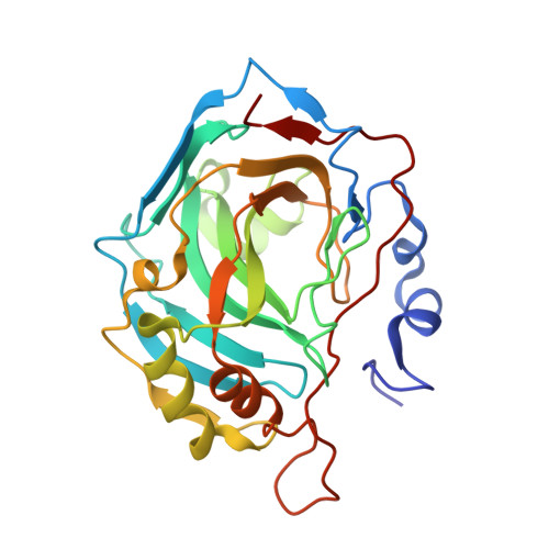
    3IBI, 3IBL, 3IBN, 3IBU

  • PubMed Abstract: 

    Two approaches were used to design inhibitors of the metalloenzyme carbonic anhydrase (CA, EC 4.2.1.1): the tail and the ring approaches. Aliphatic sulfamates constitute a class of CA inhibitors (CAIs) that cannot be classified in either one of these categories. We report here the detailed inhibition profile of four such compounds against isoforms CAs I-XIV, the first crystallographic structures of these compounds in adduct with isoform II, and molecular modeling studies for their interaction with hCA IX. Aliphatic monosulfamates/bis-sulfamates were nanomolar inhibitors of hCAs II, IX, and XII, unlike aromatic/heterocyclic sulfonamides that promiscuously inhibit most CA isozymes with low nanomolar affinity. The bis-sulfamates incorporating 8 or 10 carbon atoms showed higher affinity for the tumor-associated hCA IX compared to hCA II, whereas the opposite was true for the monosulfamates. The explanation for their interaction with CA active site furnishes insights for obtaining compounds with increased affinity/selectivity for various isozymes.


  • Organizational Affiliation
    • Istituto di Chimica Biomolecolare-CNR, Pozzuoli, Italy.

Macromolecule Content 

  • Total Structure Weight: 29.93 kDa 
  • Atom Count: 2,272 
  • Modeled Residue Count: 258 
  • Deposited Residue Count: 259 
  • Unique protein chains: 1

Macromolecules

Find similar proteins by:|  3D Structure
Entity ID: 1
MoleculeChains  Sequence LengthOrganismDetailsImage
Carbonic anhydrase 2259Homo sapiensMutation(s): 0 
EC: 4.2.1.1 (PDB Primary Data), 4.2.1.69 (UniProt)
UniProt & NIH Common Fund Data Resources
Find proteins for P00918 (Homo sapiens)
Explore P00918 
Go to UniProtKB:  P00918
PHAROS:  P00918
GTEx:  ENSG00000104267 
Entity Groups
Sequence Clusters30% Identity50% Identity70% Identity90% Identity95% Identity100% Identity
UniProt GroupP00918
Sequence Annotations
Expand
Reference Sequence

Experimental Data & Validation

Experimental Data

  • Method: X-RAY DIFFRACTION
  • Resolution: 2.20 Å
  • R-Value Free:  0.223 (Depositor), 0.214 (DCC) 
  • R-Value Work:  0.187 (Depositor), 0.176 (DCC) 
Space Group: P 1 21 1
Unit Cell:
Length ( Å )Angle ( ˚ )
a = 41.99α = 90
b = 41.34β = 104.25
c = 71.85γ = 90
Software Package:
Software NamePurpose
CNSrefinement
DENZOdata reduction
SCALEPACKdata scaling
CNSphasing

Structure Validation

View Full Validation Report


Ligand Structure Quality Assessment 


Entry History 

Deposition Data

Revision History  (Full details and data files)

  • Version 1.0: 2009-09-22
    Type: Initial release
  • Version 1.1: 2011-07-13
    Changes: Version format compliance
  • Version 1.2: 2011-08-24
    Changes: Atomic model, Database references, Structure summary
  • Version 1.3: 2023-11-01
    Changes: Data collection, Database references, Derived calculations, Refinement description