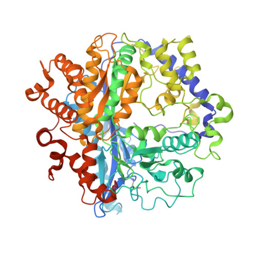
Mechanistic details of glutathione biosynthesis revealed by crystal structures of Saccharomyces cerevisiae glutamate cysteine ligase.
Biterova, E.I., Barycki, J.J.(2009) J Biological Chem 284: 32700-32708
- PubMed: 19726687
- DOI: https://doi.org/10.1074/jbc.M109.025114
- Primary Citation of Related Structures:
3IG5, 3IG8 - PubMed Abstract:
Glutathione is a thiol-disulfide exchange peptide critical for buffering oxidative or chemical stress, and an essential cofactor in several biosynthesis and detoxification pathways. The rate-limiting step in its de novo biosynthesis is catalyzed by glutamate cysteine ligase, a broadly expressed enzyme for which limited structural information is available in higher eukaryotic species. Structural data are critical to the understanding of clinical glutathione deficiency, as well as rational design of enzyme modulators that could impact human disease progression. Here, we have determined the structures of Saccharomyces cerevisiae glutamate cysteine ligase (ScGCL) in the presence of glutamate and MgCl(2) (2.1 A; R = 18.2%, R(free) = 21.9%), and in complex with glutamate, MgCl(2), and ADP (2.7 A; R = 19.0%, R(free) = 24.2%). Inspection of these structures reveals an unusual binding pocket for the alpha-carboxylate of the glutamate substrate and an ATP-independent Mg(2+) coordination site, clarifying the Mg(2+) dependence of the enzymatic reaction. The ScGCL structures were further used to generate a credible homology model of the catalytic subunit of human glutamate cysteine ligase (hGCLC). Examination of the hGCLC model suggests that post-translational modifications of cysteine residues may be involved in the regulation of enzymatic activity, and elucidates the molecular basis of glutathione deficiency associated with patient hGCLC mutations.
- Department of Biochemistry and the Redox Biology Center, University of Nebraska, Lincoln, Nebraska 68588-0664, USA.
Organizational Affiliation:


