3IGU

Crystal structure of human alpha-N-acetylgalactosaminidase, covalent intermediate


Experimental Data Snapshot

  • Method: X-RAY DIFFRACTION
  • Resolution: 2.15 Å
  • R-Value Free: 
    0.199 (Depositor), 0.210 (DCC) 
  • R-Value Work: 
    0.164 (Depositor), 0.170 (DCC) 
  • R-Value Observed: 
    0.166 (Depositor) 

Starting Model: experimental
View more details

wwPDB Validation   3D Report Full Report


Ligand Structure Quality Assessment 

Created with Raphaël 2.3.0Worse 01 BetterLigand structure goodness of fit to experimental dataBest fitted NAGClick on this verticalbar to view detailsBest fitted 7JZClick on this verticalbar to view details

This is version 2.3 of the entry. See complete history


Literature

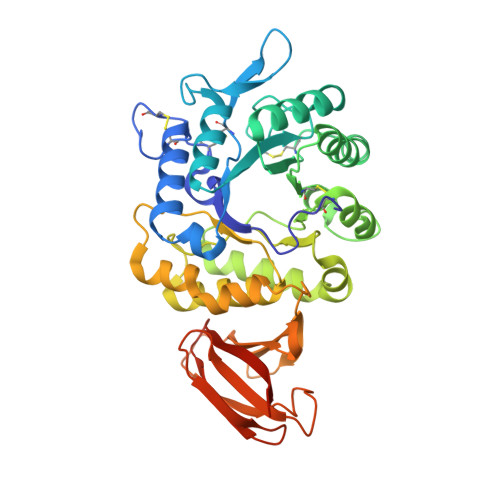
The 1.9 a structure of human alpha-N-acetylgalactosaminidase: The molecular basis of Schindler and Kanzaki diseases

Clark, N.E.Garman, S.C.

(2009) J Mol Biology 393: 435-447

  • DOI: https://doi.org/10.1016/j.jmb.2009.08.021
  • Primary Citation of Related Structures:  
    3H53, 3H54, 3H55, 3IGU

  • PubMed Abstract: 

    alpha-N-acetylgalactosaminidase (alpha-NAGAL; E.C. 3.2.1.49) is a lysosomal exoglycosidase that cleaves terminal alpha-N-acetylgalactosamine residues from glycopeptides and glycolipids. In humans, a deficiency of alpha-NAGAL activity results in the lysosomal storage disorders Schindler disease and Kanzaki disease. To better understand the molecular defects in the diseases, we determined the crystal structure of human alpha-NAGAL after expressing wild-type and glycosylation-deficient glycoproteins in recombinant insect cell expression systems. We measured the enzymatic parameters of our purified wild-type and mutant enzymes, establishing their enzymatic equivalence. To investigate the binding specificity and catalytic mechanism of the human alpha-NAGAL enzyme, we determined three crystallographic complexes with different catalytic products bound in the active site of the enzyme. To better understand how individual defects in the alpha-NAGAL glycoprotein lead to Schindler disease, we analyzed the effect of disease-causing mutations on the three-dimensional structure.


  • Organizational Affiliation

    Department of Biochemistry and Molecular Biology, University of Massachusetts, Amherst, 01003, USA.


Macromolecules
Find similar proteins by:  (by identity cutoff)  |  3D Structure
Entity ID: 1
MoleculeChains Sequence LengthOrganismDetailsImage
Alpha-N-acetylgalactosaminidase
A, B
400Homo sapiensMutation(s): 1 
Gene Names: NAGA
EC: 3.2.1.49
UniProt & NIH Common Fund Data Resources
Find proteins for P17050 (Homo sapiens)
Explore P17050 
Go to UniProtKB:  P17050
PHAROS:  P17050
GTEx:  ENSG00000198951 
Entity Groups  
Sequence Clusters30% Identity50% Identity70% Identity90% Identity95% Identity100% Identity
UniProt GroupP17050
Glycosylation
Glycosylation Sites: 3Go to GlyGen: P17050-1
Sequence Annotations
Expand
  • Reference Sequence
Oligosaccharides

Help

Entity ID: 2
MoleculeChains Length2D Diagram Glycosylation3D Interactions
2-acetamido-2-deoxy-beta-D-glucopyranose-(1-4)-2-acetamido-2-deoxy-beta-D-glucopyranose
C
2N-Glycosylation
Glycosylation Resources
GlyTouCan:  G42666HT
GlyCosmos:  G42666HT
GlyGen:  G42666HT
Entity ID: 3
MoleculeChains Length2D Diagram Glycosylation3D Interactions
alpha-D-mannopyranose-(1-3)-[alpha-D-mannopyranose-(1-6)]beta-D-mannopyranose-(1-4)-2-acetamido-2-deoxy-beta-D-glucopyranose-(1-4)-2-acetamido-2-deoxy-beta-D-glucopyranose
D, F
5N-Glycosylation
Glycosylation Resources
GlyTouCan:  G22768VO
GlyCosmos:  G22768VO
GlyGen:  G22768VO
Entity ID: 4
MoleculeChains Length2D Diagram Glycosylation3D Interactions
2-acetamido-2-deoxy-beta-D-glucopyranose-(1-4)-[alpha-L-fucopyranose-(1-6)]2-acetamido-2-deoxy-beta-D-glucopyranose
E
3N-Glycosylation
Glycosylation Resources
GlyTouCan:  G21290RB
GlyCosmos:  G21290RB
GlyGen:  G21290RB
Small Molecules
Ligands 3 Unique
IDChains Name / Formula / InChI Key2D Diagram3D Interactions
NAG
Query on NAG

Download Ideal Coordinates CCD File 
R [auth A],
Y [auth B]
2-acetamido-2-deoxy-beta-D-glucopyranose
C8 H15 N O6
OVRNDRQMDRJTHS-FMDGEEDCSA-N
7JZ
Query on 7JZ

Download Ideal Coordinates CCD File 
Q [auth A],
X [auth B]
2-deoxy-2,2-difluoro-beta-D-lyxo-hexopyranose
C6 H10 F2 O5
SHBCJQOMZTUYEL-MGCNEYSASA-N
GOL
Query on GOL

Download Ideal Coordinates CCD File 
G [auth A]
H [auth A]
I [auth A]
J [auth A]
K [auth A]
GLYCEROL
C3 H8 O3
PEDCQBHIVMGVHV-UHFFFAOYSA-N
Experimental Data & Validation

Experimental Data

  • Method: X-RAY DIFFRACTION
  • Resolution: 2.15 Å
  • R-Value Free:  0.199 (Depositor), 0.210 (DCC) 
  • R-Value Work:  0.164 (Depositor), 0.170 (DCC) 
  • R-Value Observed: 0.166 (Depositor) 
Space Group: C 1 2 1
Unit Cell:
Length ( Å )Angle ( ˚ )
a = 151.612α = 90
b = 113.649β = 96.14
c = 68.415γ = 90
Software Package:
Software NamePurpose
DENZOdata reduction
SCALEPACKdata scaling
REFMACrefinement
PDB_EXTRACTdata extraction
HKL-2000data reduction

Structure Validation

View Full Validation Report


Ligand Structure Quality Assessment 

Created with Raphaël 2.3.0Worse 01 BetterLigand structure goodness of fit to experimental dataBest fitted NAGClick on this verticalbar to view detailsBest fitted 7JZClick on this verticalbar to view details

Entry History 

Deposition Data

Revision History  (Full details and data files)

  • Version 1.0: 2009-10-20
    Type: Initial release
  • Version 1.1: 2011-07-13
    Changes: Advisory, Version format compliance
  • Version 1.2: 2017-11-01
    Changes: Refinement description
  • Version 2.0: 2020-07-29
    Type: Remediation
    Reason: Carbohydrate remediation
    Changes: Advisory, Atomic model, Data collection, Database references, Derived calculations, Structure summary
  • Version 2.1: 2021-10-13
    Changes: Database references, Structure summary
  • Version 2.2: 2023-09-06
    Changes: Data collection, Refinement description
  • Version 2.3: 2024-10-16
    Changes: Structure summary