3JZ2 | pdb_00003jz2

Crystal structure of human thrombin mutant N143P in E* form


Experimental Data Snapshot

  • Method: X-RAY DIFFRACTION
  • Resolution: 2.40 Å
  • R-Value Free: 
    0.246 (Depositor), 0.260 (DCC) 
  • R-Value Work: 
    0.189 (Depositor), 0.200 (DCC) 
  • R-Value Observed: 
    0.192 (Depositor) 

Starting Model: experimental
View more details

wwPDB Validation   3D Report Full Report


Ligand Structure Quality Assessment 

Created with Raphaël 2.3.0Worse 01 BetterLigand structure goodness of fit to experimental dataBest fitted NAGClick on this verticalbar to view details

This is version 1.5 of the entry. See complete history


Literature

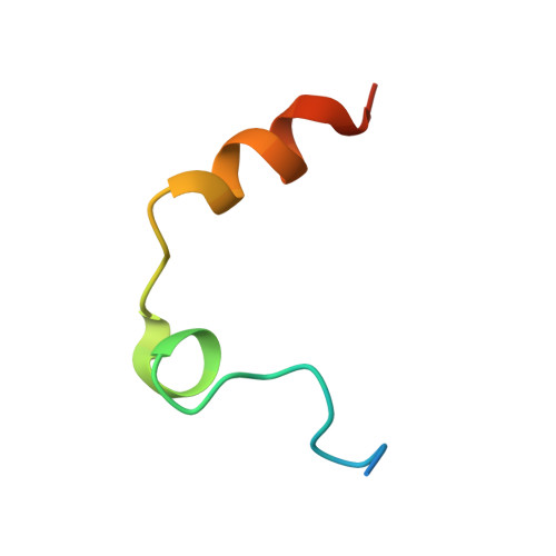
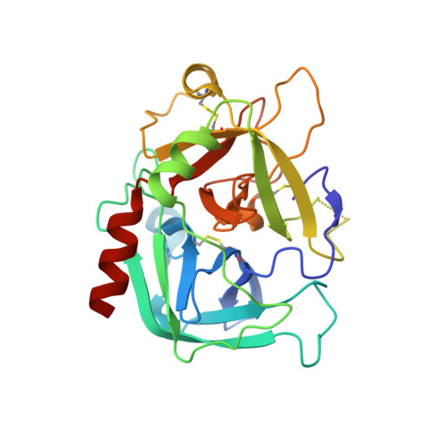
Mutant N143P reveals how Na+ activates thrombin

Niu, W.Chen, Z.Bush-Pelc, L.A.Bah, A.Gandhi, P.S.Di Cera, E.

(2009) J Biological Chem 284: 36175-36185

  • DOI: https://doi.org/10.1074/jbc.M109.069500
  • Primary Citation of Related Structures:  
    3JZ1, 3JZ2

  • PubMed Abstract: 

    The molecular mechanism of thrombin activation by Na(+) remains elusive. Its kinetic formulation requires extension of the classical Botts-Morales theory for the action of a modifier on an enzyme to correctly account for the contribution of the E*, E, and E:Na(+) forms. The extended scheme establishes that analysis of k(cat) unequivocally identifies allosteric transduction of Na(+) binding into enhanced catalytic activity. The thrombin mutant N143P features no Na(+)-dependent enhancement of k(cat) yet binds Na(+) with an affinity comparable to that of wild type. Crystal structures of the mutant in the presence and absence of Na(+) confirm that Pro(143) abrogates the important H-bond between the backbone N atom of residue 143 and the carbonyl O atom of Glu(192), which in turn controls the orientation of the Glu(192)-Gly(193) peptide bond and the correct architecture of the oxyanion hole. We conclude that Na(+) activates thrombin by securing the correct orientation of the Glu(192)-Gly(193) peptide bond, which is likely flipped in the absence of cation. Absolute conservation of the 143-192 H-bond in trypsin-like proteases and the importance of the oxyanion hole in protease function suggest that this mechanism of Na(+) activation is present in all Na(+)-activated trypsin-like proteases.


  • Organizational Affiliation

    Department of Biochemistry and Molecular Biophysics, Washington University School of Medicine, St. Louis, Missouri 63110.


Macromolecules
Find similar proteins by:  (by identity cutoff)  |  3D Structure
Entity ID: 1
MoleculeChains Sequence LengthOrganismDetailsImage
Thrombin light chain36Homo sapiensMutation(s): 0 
Gene Names: F2
EC: 3.4.21.5
UniProt & NIH Common Fund Data Resources
Find proteins for P00734 (Homo sapiens)
Explore P00734 
Go to UniProtKB:  P00734
PHAROS:  P00734
GTEx:  ENSG00000180210 
Entity Groups  
Sequence Clusters30% Identity50% Identity70% Identity90% Identity95% Identity100% Identity
UniProt GroupP00734
Sequence Annotations
Expand
  • Reference Sequence
Find similar proteins by:  (by identity cutoff)  |  3D Structure
Entity ID: 2
MoleculeChains Sequence LengthOrganismDetailsImage
Thrombin heavy chain259Homo sapiensMutation(s): 1 
Gene Names: F2
EC: 3.4.21.5
UniProt & NIH Common Fund Data Resources
Find proteins for P00734 (Homo sapiens)
Explore P00734 
Go to UniProtKB:  P00734
PHAROS:  P00734
GTEx:  ENSG00000180210 
Entity Groups  
Sequence Clusters30% Identity50% Identity70% Identity90% Identity95% Identity100% Identity
UniProt GroupP00734
Glycosylation
Glycosylation Sites: 1Go to GlyGen: P00734-1
Sequence Annotations
Expand
  • Reference Sequence
Experimental Data & Validation

Experimental Data

  • Method: X-RAY DIFFRACTION
  • Resolution: 2.40 Å
  • R-Value Free:  0.246 (Depositor), 0.260 (DCC) 
  • R-Value Work:  0.189 (Depositor), 0.200 (DCC) 
  • R-Value Observed: 0.192 (Depositor) 
Space Group: P 43
Unit Cell:
Length ( Å )Angle ( ˚ )
a = 57.89α = 90
b = 57.89β = 90
c = 119.77γ = 90
Software Package:
Software NamePurpose
MAR345dtbdata collection
MOLREPphasing
REFMACrefinement
HKL-2000data reduction
HKL-2000data scaling

Structure Validation

View Full Validation Report


Ligand Structure Quality Assessment 

Created with Raphaël 2.3.0Worse 01 BetterLigand structure goodness of fit to experimental dataBest fitted NAGClick on this verticalbar to view details

Entry History 

Deposition Data

Revision History  (Full details and data files)

  • Version 1.0: 2009-10-20
    Type: Initial release
  • Version 1.1: 2011-07-13
    Changes: Non-polymer description, Version format compliance
  • Version 1.2: 2020-07-29
    Type: Remediation
    Reason: Carbohydrate remediation
    Changes: Data collection, Derived calculations, Structure summary
  • Version 1.3: 2021-10-13
    Changes: Database references, Structure summary
  • Version 1.4: 2023-09-06
    Changes: Data collection, Refinement description
  • Version 1.5: 2024-11-06
    Changes: Structure summary