3KK1

HIV-1 reverse transcriptase-DNA complex with nuceotide inhibitor GS-9148-diphosphate bound in nucleotide site


Experimental Data Snapshot

  • Method: X-RAY DIFFRACTION
  • Resolution: 2.70 Å
  • R-Value Free: 
    0.274 (Depositor), 0.270 (DCC) 
  • R-Value Work: 
    0.204 (Depositor), 0.200 (DCC) 
  • R-Value Observed: 
    0.208 (Depositor) 

Starting Model: experimental
View more details

wwPDB Validation   3D Report Full Report


Ligand Structure Quality Assessment 

Created with Raphaël 2.3.0Worse 01 BetterLigand structure goodness of fit to experimental dataBest fitted 914Click on this verticalbar to view details

This is version 1.3 of the entry. See complete history


Literature

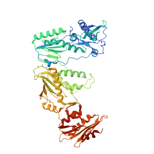
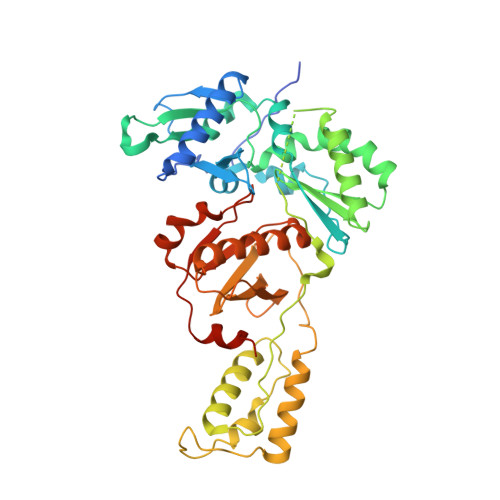
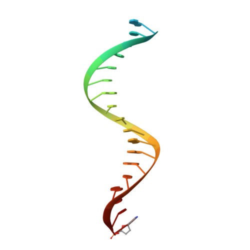
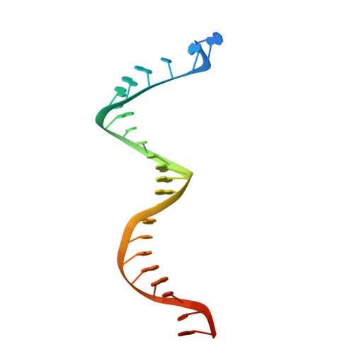
Visualizing the molecular interactions of a nucleotide analog, GS-9148, with HIV-1 reverse transcriptase-DNA complex.

Lansdon, E.B.Samuel, D.Lagpacan, L.Brendza, K.M.White, K.L.Hung, M.Liu, X.Boojamra, C.G.Mackman, R.L.Cihlar, T.Ray, A.S.McGrath, M.E.Swaminathan, S.

(2010) J Mol Biol 397: 967-978

  • DOI: https://doi.org/10.1016/j.jmb.2010.02.019
  • Primary Citation of Related Structures:  
    3KJV, 3KK1, 3KK2, 3KK3

  • PubMed Abstract: 

    GS-9148 ([5-(6-amino-purin-9-yl)-4-fluoro-2,5-dihydro-furan-2-yloxymethyl]-phosphonic acid) is a dAMP (2'-deoxyadenosine monophosphate) analog that maintains its antiviral activity against drug-resistant HIV. Crystal structures for HIV-1 reverse transcriptase (RT) bound to double-stranded DNA, ternary complexes with either GS-9148-diphosphate or 2'-deoxyadenosine triphosphate (dATP), and a post-incorporation structure with GS-9148 translocated to the priming site were obtained to gain insight into the mechanism of RT inhibition. The binding of either GS-9148-diphosphate or dATP to the binary RT-DNA complex resulted in the fingers subdomain closing around the incoming substrate. This produced up to a 9 A shift in the tips of the fingers subdomain as it closed toward the palm and thumb subdomains. GS-9148-diphosphate shows a similar binding mode as dATP in the nucleotide-binding site. Residues whose mutations confer resistance to nucleotide/nucleoside RT inhibitors, such as M184, Y115, L74, and K65, show little to no shift in orientation whether GS-9148-diphosphate or dATP is bound. One difference observed in binding is the position of the central ring. The dihydrofuran ring of GS-9148-diphosphate interacts with the aromatic side chain of Y115 more than does the ribose ring of dATP, possibly picking up a favorable pi-pi interaction. The ability of GS-9148-diphosphate to mimic the active-site contacts of dATP may explain its effective inhibition of RT and maintained activity against resistance mutations. Interestingly, the 2'-fluoro moiety of GS-9148-diphosphate was found in close proximity to the Q151 side chain, potentially explaining the observed moderately reduced susceptibly to GS-9148 conferred by Q151M mutation.


  • Organizational Affiliation

    Gilead Sciences, Inc., 333 Lakeside Drive, Foster City, CA 94404, USA. eric.lansdon@gilead.com


Macromolecules

Find similar proteins by:  (by identity cutoff)  |  3D Structure
Entity ID: 1
MoleculeChains Sequence LengthOrganismDetailsImage
Reverse transcriptase p66 subunit560HIV-1 M:B_HXB2RMutation(s): 2 
Gene Names: gag-pol
EC: 2.7.7.49
UniProt
Find proteins for P04585 (Human immunodeficiency virus type 1 group M subtype B (isolate HXB2))
Explore P04585 
Go to UniProtKB:  P04585
Entity Groups  
Sequence Clusters30% Identity50% Identity70% Identity90% Identity95% Identity100% Identity
UniProt GroupP04585
Sequence Annotations
Expand
  • Reference Sequence
Find similar proteins by:  (by identity cutoff)  |  3D Structure
Entity ID: 2
MoleculeChains Sequence LengthOrganismDetailsImage
Reverse transcriptase p51 subunit452HIV-1 M:B_HXB2RMutation(s): 1 
Gene Names: gag-pol
EC: 2.7.7.49
UniProt
Find proteins for P04585 (Human immunodeficiency virus type 1 group M subtype B (isolate HXB2))
Explore P04585 
Go to UniProtKB:  P04585
Entity Groups  
Sequence Clusters30% Identity50% Identity70% Identity90% Identity95% Identity100% Identity
UniProt GroupP04585
Sequence Annotations
Expand
  • Reference Sequence
Small Molecules
Ligands 3 Unique
IDChains Name / Formula / InChI Key2D Diagram3D Interactions
914
Query on 914

Download Ideal Coordinates CCD File 
I [auth A][(2R,5R)-5-(6-aminopurin-9-yl)-4-fluoro-2,5-dihydrofuran-2-yl]oxymethyl-[hydroxy(phosphonooxy)phosphoryl]oxy-phosphinic acid
C10 H13 F N5 O11 P3
VWRYHMYAWKXIKG-QUBYGPBYSA-N
SO4
Query on SO4

Download Ideal Coordinates CCD File 
E [auth A],
F [auth A],
J [auth P]
SULFATE ION
O4 S
QAOWNCQODCNURD-UHFFFAOYSA-L
MG
Query on MG

Download Ideal Coordinates CCD File 
G [auth A],
H [auth A]
MAGNESIUM ION
Mg
JLVVSXFLKOJNIY-UHFFFAOYSA-N
Binding Affinity Annotations 
IDSourceBinding Affinity
914 BindingDB:  3KK1 IC50: 1900 (nM) from 1 assay(s)
Experimental Data & Validation

Experimental Data

  • Method: X-RAY DIFFRACTION
  • Resolution: 2.70 Å
  • R-Value Free:  0.274 (Depositor), 0.270 (DCC) 
  • R-Value Work:  0.204 (Depositor), 0.200 (DCC) 
  • R-Value Observed: 0.208 (Depositor) 
Space Group: C 2 2 21
Unit Cell:
Length ( Å )Angle ( ˚ )
a = 165.753α = 90
b = 169.239β = 90
c = 103.026γ = 90
Software Package:
Software NamePurpose
BOSdata collection
EPMRphasing
CNXrefinement
HKL-2000data reduction
HKL-2000data scaling

Structure Validation

View Full Validation Report


Ligand Structure Quality Assessment 

Created with Raphaël 2.3.0Worse 01 BetterLigand structure goodness of fit to experimental dataBest fitted 914Click on this verticalbar to view details

Entry History 

Deposition Data

Revision History  (Full details and data files)

  • Version 1.0: 2010-03-23
    Type: Initial release
  • Version 1.1: 2011-07-13
    Changes: Version format compliance
  • Version 1.2: 2021-10-13
    Changes: Database references, Derived calculations
  • Version 1.3: 2023-09-06
    Changes: Data collection, Refinement description