3KNE | pdb_00003kne

Carbonic Anhydrase II H64C mutant in complex with an in situ formed triazole


Experimental Data Snapshot

  • Method: X-RAY DIFFRACTION
  • Resolution: 1.35 Å
  • R-Value Free: 
    0.183 (Depositor), 0.151 (DCC) 
  • R-Value Work: 
    0.139 (DCC) 
  • R-Value Observed: 
    0.128 (Depositor) 

wwPDB Validation 3D Report Full Report

Validation slider image for 3KNE

Ligand Structure Quality Assessment 


This is version 1.5 of the entry. See complete history

Literature

Stereo- and Regioselective Azide/Alkyne Cycloadditions in Carbonic Anhydrase II via Tethering, Monitored by Crystallography and Mass Spectrometry.

Schulze Wischeler, J.Sun, D.Sandner, N.U.Linne, U.Heine, A.Koert, U.Klebe, G.

(2011) Chemistry 17: 5842-5851

  • DOI: https://doi.org/10.1002/chem.201002437
  • Primary Citation Related Structures: 
    3KIG, 3KNE

  • PubMed Abstract: 

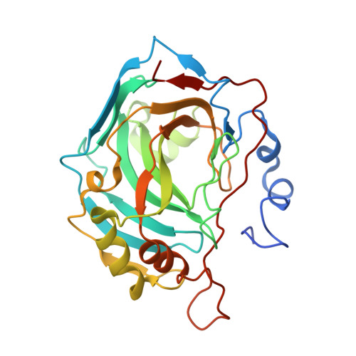
    The carbonic anhydrase II mutant His64Cys was prepared and applied to tethered alkyne/azide cycloaddition reactions. The azide component could be tethered to the enzyme surface through a disulfide bridge, while the alkyne component was reversibly coordinated through a sulfonamide anchor to the zinc ion in the original catalytic center of the enzyme. The incipient orientation of the reactants in the binding site and of the formed triazole product were characterized by crystallography. The reaction progression could be monitored by HPLC-MS analysis.


  • Organizational Affiliation
    • Institut für Pharmazeutische Chemie, Philipps-Universität Marburg, Marbacher Weg 6, 35032 Marburg, Germany.

Macromolecule Content 

  • Total Structure Weight: 30.68 kDa 
  • Atom Count: 2,331 
  • Modeled Residue Count: 257 
  • Deposited Residue Count: 265 
  • Unique protein chains: 1

Macromolecules

Find similar proteins by:|  3D Structure
Entity ID: 1
MoleculeChains  Sequence LengthOrganismDetailsImage
Carbonic anhydrase 2265Homo sapiensMutation(s): 1 
EC: 4.2.1.1 (PDB Primary Data), 4.2.1.69 (UniProt)
UniProt & NIH Common Fund Data Resources
Find proteins for P00918 (Homo sapiens)
Explore P00918 
Go to UniProtKB:  P00918
PHAROS:  P00918
GTEx:  ENSG00000104267 
Entity Groups
Sequence Clusters30% Identity50% Identity70% Identity90% Identity95% Identity100% Identity
UniProt GroupP00918
Sequence Annotations
Expand
Reference Sequence

Small Molecules

Ligands 3 Unique
IDChains Name / Formula / InChI Key2D Diagram3D Interactions
DAW

Query on DAW


Download:Ideal Coordinates CCD File
D [auth A]N-[(S)-(1-{2-oxo-2-[(3-sulfanylpropyl)amino]ethyl}-1H-1,2,3-triazol-5-yl)(phenyl)methyl]-4-sulfamoylbenzamide
C21 H24 N6 O4 S2
SQLGLXFRIDQRKD-FQEVSTJZSA-N
BE7

Query on BE7


Download:Ideal Coordinates CCD File
C [auth A](4-CARBOXYPHENYL)(CHLORO)MERCURY
C7 H5 Cl Hg O2
YFZOUMNUDGGHIW-UHFFFAOYSA-M
ZN

Query on ZN


Download:Ideal Coordinates CCD File
B [auth A]ZINC ION
Zn
PTFCDOFLOPIGGS-UHFFFAOYSA-N

Experimental Data & Validation

Experimental Data

  • Method: X-RAY DIFFRACTION
  • Resolution: 1.35 Å
  • R-Value Free:  0.183 (Depositor), 0.151 (DCC) 
  • R-Value Work:  0.139 (DCC) 
  • R-Value Observed: 0.128 (Depositor) 
Space Group: P 1 21 1
Unit Cell:
Length ( Å )Angle ( ˚ )
a = 42.4α = 90
b = 41.5β = 104.3
c = 72γ = 90
Software Package:
Software NamePurpose
MAR345data collection
SHELXmodel building
SHELXL-97refinement
HKL-2000data reduction
HKL-2000data scaling
SHELXphasing

Structure Validation

View Full Validation Report


Ligand Structure Quality Assessment 


Entry History 

Deposition Data

Revision History  (Full details and data files)

  • Version 1.0: 2011-05-18
    Type: Initial release
  • Version 1.1: 2011-07-13
    Changes: Version format compliance
  • Version 1.2: 2017-11-01
    Changes: Refinement description
  • Version 1.3: 2021-10-13
    Changes: Database references, Derived calculations
  • Version 1.4: 2024-02-28
    Changes: Data collection, Structure summary
  • Version 1.5: 2024-10-16
    Changes: Structure summary