3L2Y | pdb_00003l2y

The structure of C-reactive protein bound to phosphoethanolamine


Experimental Data Snapshot

  • Method: X-RAY DIFFRACTION
  • Resolution: 2.70 Å
  • R-Value Free: 
    0.246 (Depositor), 0.235 (DCC) 
  • R-Value Work: 
    0.224 (Depositor), 0.215 (DCC) 
  • R-Value Observed: 
    0.225 (Depositor) 

wwPDB Validation 3D Report Full Report

Validation slider image for 3L2Y

This is version 1.2 of the entry. See complete history

Literature

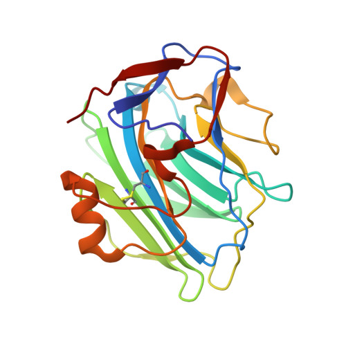
Structural basis of ligand specificity in the human pentraxins, C-reactive protein and serum amyloid P component.

Mikolajek, H.Kolstoe, S.E.Pye, V.E.Mangione, P.Pepys, M.B.Wood, S.P.

(2011) J Mol Recognit 24: 371-377

  • DOI: https://doi.org/10.1002/jmr.1090
  • Primary Citation Related Structures: 
    3KQR, 3L2Y

  • PubMed Abstract: 

    The normal physiological roles of the phylogenetically conserved human plasma proteins C-reactive protein (CRP) and serum amyloid P component (SAP) are not known. Novel drugs targeting their ligand specificities are in clinical development as both proteins have significant pathophysiological effects, SAP in promoting amyloidosis and CRP in exacerbating ischemic injury. Both proteins bind to phosphoethanolamine and we show here that, under physiological conditions, phosphoethanolamine is bound with higher affinity by human SAP than by human CRP. An explanation is provided by X-ray crystal structures that show SAP residue Tyr74 allowing additional hydrophobic protein-ligand interactions compared with the equivalent Thr76 of CRP. Docking simulations show many more low energy positions for phosphoethanolamine bound by CRP than by SAP and are consistent with the crystallographic and functional binding results. These fundamental observations on structure-activity relationships will aid the design of improved pentraxin targeting drugs.


  • Organizational Affiliation
    • Laboratory of Protein Crystallography, Acute Phase Proteins, Division of Medicine, Royal Free Campus, University College London Medical School, Rowland Hill Street, London NW3 2PF, UK.

Macromolecule Content 

  • Total Structure Weight: 465.79 kDa 
  • Atom Count: 32,993 
  • Modeled Residue Count: 4,120 
  • Deposited Residue Count: 4,120 
  • Unique protein chains: 1

Macromolecules

Find similar proteins by:|  3D Structure
Entity ID: 1
MoleculeChains  Sequence LengthOrganismDetailsImage
C-reactive protein
A, B, C, D, E
A, B, C, D, E, F, G, H, I, J, K, L, M, N, O, P, Q, R, S, T
206Homo sapiensMutation(s): 0 
UniProt & NIH Common Fund Data Resources
Find proteins for P02741 (Homo sapiens)
Explore P02741 
Go to UniProtKB:  P02741
PHAROS:  P02741
GTEx:  ENSG00000132693 
Entity Groups
Sequence Clusters30% Identity50% Identity70% Identity90% Identity95% Identity100% Identity
UniProt GroupP02741
Sequence Annotations
Expand
Reference Sequence

Small Molecules

Ligands 2 Unique
IDChains Name / Formula / InChI Key2D Diagram3D Interactions
OPE

Query on OPE


Download:Ideal Coordinates CCD File
AA [auth C]
BB [auth L]
DA [auth D]
EB [auth M]
GA [auth E]
AA [auth C],
BB [auth L],
DA [auth D],
EB [auth M],
GA [auth E],
HB [auth N],
JA [auth F],
KB [auth O],
MA [auth G],
NB [auth P],
PA [auth H],
QB [auth Q],
SA [auth I],
TB [auth R],
VA [auth J],
W [auth A],
WB [auth S],
X [auth B],
YA [auth K],
ZB [auth T]
PHOSPHORIC ACID MONO-(2-AMINO-ETHYL) ESTER
C2 H8 N O4 P
SUHOOTKUPISOBE-UHFFFAOYSA-N
CA

Query on CA


Download:Ideal Coordinates CCD File
AB [auth K]
AC [auth T]
BA [auth C]
BC [auth T]
CA [auth C]
AB [auth K],
AC [auth T],
BA [auth C],
BC [auth T],
CA [auth C],
CB [auth L],
DB [auth L],
EA [auth D],
FA [auth D],
FB [auth M],
GB [auth M],
HA [auth E],
IA [auth E],
IB [auth N],
JB [auth N],
KA [auth F],
LA [auth F],
LB [auth O],
MB [auth O],
NA [auth G],
OA [auth G],
OB [auth P],
PB [auth P],
QA [auth H],
RA [auth H],
RB [auth Q],
SB [auth Q],
TA [auth I],
U [auth A],
UA [auth I],
UB [auth R],
V [auth A],
VB [auth R],
WA [auth J],
XA [auth J],
XB [auth S],
Y [auth B],
YB [auth S],
Z [auth B],
ZA [auth K]
CALCIUM ION
Ca
BHPQYMZQTOCNFJ-UHFFFAOYSA-N

Experimental Data & Validation

Experimental Data

  • Method: X-RAY DIFFRACTION
  • Resolution: 2.70 Å
  • R-Value Free:  0.246 (Depositor), 0.235 (DCC) 
  • R-Value Work:  0.224 (Depositor), 0.215 (DCC) 
  • R-Value Observed: 0.225 (Depositor) 
Space Group: P 41
Unit Cell:
Length ( Å )Angle ( ˚ )
a = 278.448α = 90
b = 278.448β = 90
c = 92.111γ = 90
Software Package:
Software NamePurpose
MOSFLMdata reduction
SCALAdata scaling
PHASERphasing
PHENIXrefinement
PDB_EXTRACTdata extraction
ADSCdata collection

Structure Validation

View Full Validation Report


Entry History 

Deposition Data

Revision History  (Full details and data files)

  • Version 1.0: 2010-12-08
    Type: Initial release
  • Version 1.1: 2011-07-13
    Changes: Version format compliance
  • Version 1.2: 2025-03-26
    Changes: Data collection, Database references, Derived calculations, Refinement description, Structure summary