3L9R | pdb_00003l9r

Crystal structure of bovine CD1b3 with endogenously bound ligands


Experimental Data Snapshot

  • Method: X-RAY DIFFRACTION
  • Resolution: 2.30 Å
  • R-Value Free: 
    0.248 (Depositor), 0.260 (DCC) 
  • R-Value Work: 
    0.201 (Depositor), 0.213 (DCC) 
  • R-Value Observed: 
    0.203 (Depositor) 

Starting Model: experimental
View more details

wwPDB Validation 3D Report Full Report

Validation slider image for 3L9R

Ligand Structure Quality Assessment 


This is version 2.2 of the entry. See complete history

Literature

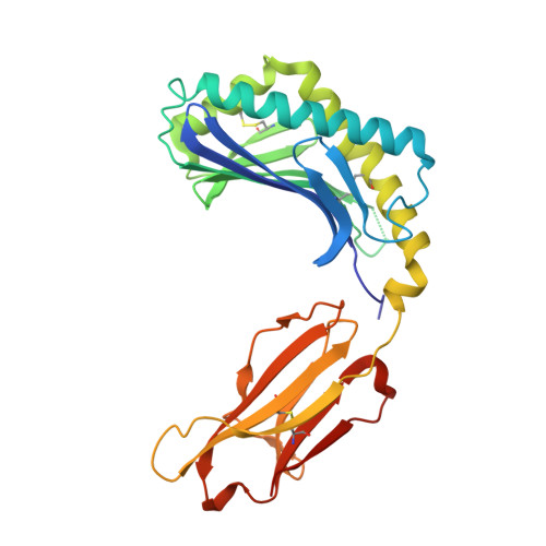
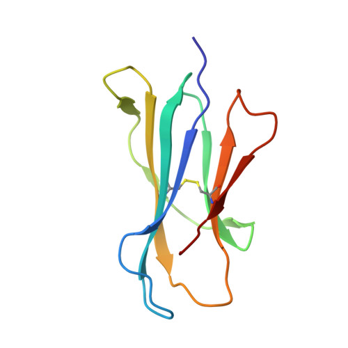
Crystal structure of bovine CD1b3 with endogenously bound ligands.

Girardi, E.Wang, J.Mac, T.T.Versluis, C.Bhowruth, V.Besra, G.Heck, A.J.Van Rhijn, I.Zajonc, D.M.

(2010) J Immunol 185: 376-386

  • DOI: https://doi.org/10.4049/jimmunol.1000042
  • Primary Citation Related Structures: 
    3L9R

  • PubMed Abstract: 

    The CD1 family of Ag-presenting molecules is able to display lipids to T cells by binding them within a hydrophobic groove connected to the protein surface. In particular, the CD1b isotype is capable of binding ligands with greatly varying alkyl chain lengths through a complex network of interconnected hydrophobic pockets. Interestingly, mycobacterial lipids such as glucose monomycolate exclusively bind to CD1b. We determined the crystal structure of one of the three expressed bovine CD1b proteins, CD1b3, in complex with endogenous ligands, identified by mass spectrometry as a mixture of phosphatidylcholine and phosphatidylethanolamine, and analyzed the ability of the protein to bind glycolipids in vitro. The structure reveals a complex binding groove architecture, similar to the human ortholog but with consequential differences. Intriguingly, in bovine CD1b3 only the A', C' and F' pockets are present, whereas the T' pocket previously described in human CD1b is closed. This different pocket conformation could affect the ability of boCD1b3 to recognize lipids with long acyl chains such as glucose monomycolate. However, even in the absence of a T' tunnel, bovine CD1b3 is able to bind mycolates from Rhodococcus ruber in vitro.


  • Organizational Affiliation
    • Division of Cell Biology, La Jolla Institute for Allergy and Immunology, La Jolla, CA, USA.

Macromolecule Content 

  • Total Structure Weight: 182.01 kDa 
  • Atom Count: 12,721 
  • Modeled Residue Count: 1,490 
  • Deposited Residue Count: 1,524 
  • Unique protein chains: 2

Macromolecules

Find similar proteins by:|  3D Structure
Entity ID: 1
MoleculeChains  Sequence LengthOrganismDetailsImage
CD1b3
A, C, E, G
283Bos taurusMutation(s): 0 
Gene Names: CD1B3
UniProt
Find proteins for Q1L1H6 (Bos taurus)
Explore Q1L1H6 
Go to UniProtKB:  Q1L1H6
Entity Groups
Sequence Clusters30% Identity50% Identity70% Identity90% Identity95% Identity100% Identity
UniProt GroupQ1L1H6
Glycosylation
Glycosylation Sites: 2
Sequence Annotations
Expand
Reference Sequence
Find similar proteins by:|  3D Structure
Entity ID: 2
MoleculeChains  Sequence LengthOrganismDetailsImage
Beta-2-microglobulin
B, D, F, H
98Bos taurusMutation(s): 0 
Gene Names: B2Mbeta-2-microglobulin
UniProt
Find proteins for P01888 (Bos taurus)
Explore P01888 
Go to UniProtKB:  P01888
Entity Groups
Sequence Clusters30% Identity50% Identity70% Identity90% Identity95% Identity100% Identity
UniProt GroupP01888
Sequence Annotations
Expand
Reference Sequence

Oligosaccharides

Help  
Entity ID: 3
MoleculeChains Length2D Diagram GlycosylationD Interactions
alpha-D-mannopyranose-(1-3)-beta-D-mannopyranose-(1-4)-2-acetamido-2-deoxy-beta-D-glucopyranose-(1-4)-[alpha-L-fucopyranose-(1-3)][alpha-L-fucopyranose-(1-6)]2-acetamido-2-deoxy-beta-D-glucopyranose
I
6N-Glycosylation
Glycosylation Resources
GlyTouCan: G17601US
GlyCosmos: G17601US
GlyGen: G17601US
Entity ID: 4
MoleculeChains Length2D Diagram GlycosylationD Interactions
beta-D-mannopyranose-(1-4)-2-acetamido-2-deoxy-beta-D-glucopyranose-(1-4)-[alpha-L-fucopyranose-(1-3)][alpha-L-fucopyranose-(1-6)]2-acetamido-2-deoxy-beta-D-glucopyranose
J, K, L
5N-Glycosylation
Glycosylation Resources
GlyTouCan: G92953ND
GlyCosmos: G92953ND
GlyGen: G92953ND

Small Molecules

Ligands 5 Unique
IDChains Name / Formula / InChI Key2D Diagram3D Interactions
L9R

Query on L9R


Download:Ideal Coordinates CCD File
N [auth A],
Y [auth E]
(2S)-3-(octadecanoyloxy)-2-[(9Z)-octadec-9-enoyloxy]propyl 2-(trimethylammonio)ethyl phosphate
C44 H86 N O8 P
ATHVAWFAEPLPPQ-FLHXZNPISA-N
L9Q

Query on L9Q


Download:Ideal Coordinates CCD File
FA [auth G],
Q [auth C]
(1S)-2-{[(S)-(2-aminoethoxy)(hydroxy)phosphoryl]oxy}-1-[(octadecanoyloxy)methyl]ethyl (9Z)-octadec-9-enoate
C41 H80 N O8 P
JQKOHRZNEOQNJE-VYCVAHKRSA-N
NAG

Query on NAG


Download:Ideal Coordinates CCD File
M [auth A],
P [auth C],
V [auth E],
W [auth E],
Z [auth G]
2-acetamido-2-deoxy-beta-D-glucopyranose
C8 H15 N O6
OVRNDRQMDRJTHS-FMDGEEDCSA-N
GOL

Query on GOL


Download:Ideal Coordinates CCD File
AA [auth G]
BA [auth G]
CA [auth G]
DA [auth G]
O [auth A]
AA [auth G],
BA [auth G],
CA [auth G],
DA [auth G],
O [auth A],
R [auth C],
S [auth C],
U [auth C],
X [auth E]
GLYCEROL
C3 H8 O3
PEDCQBHIVMGVHV-UHFFFAOYSA-N
CL

Query on CL


Download:Ideal Coordinates CCD File
EA [auth G],
T [auth C]
CHLORIDE ION
Cl
VEXZGXHMUGYJMC-UHFFFAOYSA-M

Experimental Data & Validation

Experimental Data

  • Method: X-RAY DIFFRACTION
  • Resolution: 2.30 Å
  • R-Value Free:  0.248 (Depositor), 0.260 (DCC) 
  • R-Value Work:  0.201 (Depositor), 0.213 (DCC) 
  • R-Value Observed: 0.203 (Depositor) 
Space Group: P 21 21 2
Unit Cell:
Length ( Å )Angle ( ˚ )
a = 137.533α = 90
b = 139.975β = 90
c = 111.953γ = 90
Software Package:
Software NamePurpose
HKL-2000data collection
MOLREPphasing
REFMACrefinement
HKL-2000data reduction
HKL-2000data scaling

Structure Validation

View Full Validation Report


Ligand Structure Quality Assessment 


Entry History 

Deposition Data

Revision History  (Full details and data files)

  • Version 1.0: 2010-06-16
    Type: Initial release
  • Version 1.1: 2011-07-13
    Changes: Version format compliance
  • Version 2.0: 2020-07-29
    Type: Remediation
    Reason: Carbohydrate remediation
    Changes: Atomic model, Data collection, Database references, Derived calculations, Structure summary
  • Version 2.1: 2023-09-06
    Changes: Data collection, Database references, Refinement description, Structure summary
  • Version 2.2: 2024-10-09
    Changes: Structure summary