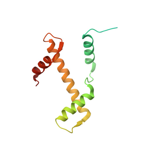
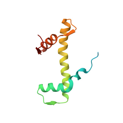
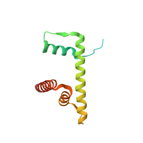
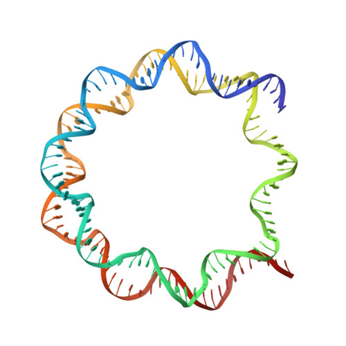
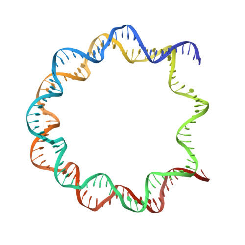
3LZ1

Crystal Structure of Nucleosome Core Particle Composed of the Widom 601 DNA Sequence (orientation 2)


Experimental Data Snapshot

  • Method: X-RAY DIFFRACTION
  • Resolution: 2.50 Å
  • R-Value Free: 0.319 
  • R-Value Work: 0.269 
  • R-Value Observed: 0.270 

Starting Model: experimental
View more details

wwPDB Validation   3D Report Full Report


This is version 1.3 of the entry. See complete history


Literature

Crystal structures of nucleosome core particles containing the '601' strong positioning sequence

Vasudevan, D.Chua, E.Y.Davey, C.A.

(2010) J Mol Biol 403: 1-10

  • DOI: https://doi.org/10.1016/j.jmb.2010.08.039
  • Primary Citation of Related Structures:  
    3LZ0, 3LZ1

  • PubMed Abstract: 

    Nucleosome positioning plays a key role in genomic regulation by defining histone-DNA context and by modulating access to specific sites. Moreover, the histone-DNA register influences the double-helix structure, which in turn can affect the association of small molecules and protein factors. Analysis of genomic and synthetic DNA has revealed sequence motifs that direct nucleosome positioning in vitro; thus, establishing the basis for the DNA sequence dependence of positioning would shed light on the mechanics of the double helix and its contribution to chromatin structure in vivo. However, acquisition of well-diffracting nucleosome core particle (NCP) crystals is extremely dependent on the DNA fragment used for assembly, and all previous NCP crystal structures have been based on human α-satellite sequences. Here, we describe the crystal structures of Xenopus NCPs containing one of the strongest known histone octamer binding and positioning sequences, the so-called '601' DNA. Two distinct 145-bp 601 crystal forms display the same histone-DNA register, which coincides with the occurrence of DNA stretching-overtwisting in both halves of the particle around five double-helical turns from the nucleosome center, giving the DNA an 'effective length' of 147 bp. As we have found previously with stretching around two turns from the nucleosome center for a centromere-based sequence, the terminal stretching observed in the 601 constructs is associated with extreme kinking into the minor groove at purine-purine (pyrimidine-pyrimidine) dinucleotide steps. In other contexts, these step types display an overall nonflexible behavior, which raises the possibility that DNA stretching in the nucleosome or extreme distortions in general have unique sequence dependency characteristics. Our findings indicate that DNA stretching is an intrinsically predisposed site-specific property of the nucleosome and suggest how NCP crystal structures with diverse DNA sequences can be obtained.


  • Organizational Affiliation

    Division of Structural and Computational Biology, School of Biological Sciences, Nanyang Technological University, 60 Nanyang Drive, Singapore 637551, Singapore.


Macromolecules

Find similar proteins by:  (by identity cutoff)  |  3D Structure
Entity ID: 1
MoleculeChains Sequence LengthOrganismDetailsImage
Histone H3.2
A, E
135Xenopus laevisMutation(s): 0 
UniProt
Find proteins for P84233 (Xenopus laevis)
Explore P84233 
Go to UniProtKB:  P84233
Entity Groups  
Sequence Clusters30% Identity50% Identity70% Identity90% Identity95% Identity100% Identity
UniProt GroupP84233
Sequence Annotations
Expand
  • Reference Sequence
Find similar proteins by:  (by identity cutoff)  |  3D Structure
Entity ID: 2
MoleculeChains Sequence LengthOrganismDetailsImage
Histone H4
B, F
102Xenopus laevisMutation(s): 0 
UniProt
Find proteins for P62799 (Xenopus laevis)
Explore P62799 
Go to UniProtKB:  P62799
Entity Groups  
Sequence Clusters30% Identity50% Identity70% Identity90% Identity95% Identity100% Identity
UniProt GroupP62799
Sequence Annotations
Expand
  • Reference Sequence
Find similar proteins by:  (by identity cutoff)  |  3D Structure
Entity ID: 3
MoleculeChains Sequence LengthOrganismDetailsImage
Histone H2A
C, G
119Xenopus laevisMutation(s): 0 
UniProt
Find proteins for Q6AZJ8 (Xenopus laevis)
Explore Q6AZJ8 
Go to UniProtKB:  Q6AZJ8
Entity Groups  
Sequence Clusters30% Identity50% Identity70% Identity90% Identity95% Identity100% Identity
UniProt GroupQ6AZJ8
Sequence Annotations
Expand
  • Reference Sequence
Find similar proteins by:  (by identity cutoff)  |  3D Structure
Entity ID: 4
MoleculeChains Sequence LengthOrganismDetailsImage
Histone H2B 1.1
D, H
125Xenopus laevisMutation(s): 0 
UniProt
Find proteins for P02281 (Xenopus laevis)
Explore P02281 
Go to UniProtKB:  P02281
Entity Groups  
Sequence Clusters30% Identity50% Identity70% Identity90% Identity95% Identity100% Identity
UniProt GroupP02281
Sequence Annotations
Expand
  • Reference Sequence
Find similar nucleic acids by:  (by identity cutoff)  |  3D Structure
Entity ID: 5
MoleculeChains LengthOrganismImage
DNA (145-MER)145N/A
Sequence Annotations
Expand
  • Reference Sequence
Find similar nucleic acids by:  (by identity cutoff)  |  3D Structure
Entity ID: 6
MoleculeChains LengthOrganismImage
DNA (145-MER)145N/A
Sequence Annotations
Expand
  • Reference Sequence
Small Molecules
Ligands 2 Unique
IDChains Name / Formula / InChI Key2D Diagram3D Interactions
MN
Query on MN

Download Ideal Coordinates CCD File 
K [auth A]
N [auth I]
O [auth I]
P [auth I]
Q [auth J]
K [auth A],
N [auth I],
O [auth I],
P [auth I],
Q [auth J],
R [auth J]
MANGANESE (II) ION
Mn
WAEMQWOKJMHJLA-UHFFFAOYSA-N
CL
Query on CL

Download Ideal Coordinates CCD File 
L [auth C],
M [auth G]
CHLORIDE ION
Cl
VEXZGXHMUGYJMC-UHFFFAOYSA-M
Experimental Data & Validation

Experimental Data

  • Method: X-RAY DIFFRACTION
  • Resolution: 2.50 Å
  • R-Value Free: 0.319 
  • R-Value Work: 0.269 
  • R-Value Observed: 0.270 
  • Space Group: P 21 21 21
Unit Cell:
Length ( Å )Angle ( ˚ )
a = 107.37α = 90
b = 109.66β = 90
c = 175.75γ = 90
Software Package:
Software NamePurpose
SCALAdata scaling
REFMACrefinement
PDB_EXTRACTdata extraction
MOSFLMdata reduction
PHASESphasing

Structure Validation

View Full Validation Report


Entry History 

Deposition Data

Revision History  (Full details and data files)

  • Version 1.0: 2010-09-15
    Type: Initial release
  • Version 1.1: 2011-07-13
    Changes: Version format compliance
  • Version 1.2: 2012-11-14
    Changes: Database references, Structure summary
  • Version 1.3: 2023-11-01
    Changes: Data collection, Database references, Derived calculations, Refinement description