3M1D | pdb_00003m1d

Structure of BIR1 from cIAP1


Experimental Data Snapshot

  • Method: X-RAY DIFFRACTION
  • Resolution: 2.00 Å
  • R-Value Free: 
    0.232 (Depositor), 0.205 (DCC) 
  • R-Value Work: 
    0.196 (Depositor) 
  • R-Value Observed: 
    0.198 (Depositor) 

Starting Model: experimental
View more details

wwPDB Validation 3D Report Full Report

Validation slider image for 3M1D

This is version 1.4 of the entry. See complete history

Literature

Asymmetric recruitment of cIAPs by TRAF2

Mace, P.D.Smits, C.Vaux, D.L.Silke, J.Day, C.L.

(2010) J Mol Biology 400: 8-15

  • DOI: https://doi.org/10.1016/j.jmb.2010.04.055
  • Primary Citation Related Structures: 
    3M1D

  • PubMed Abstract: 

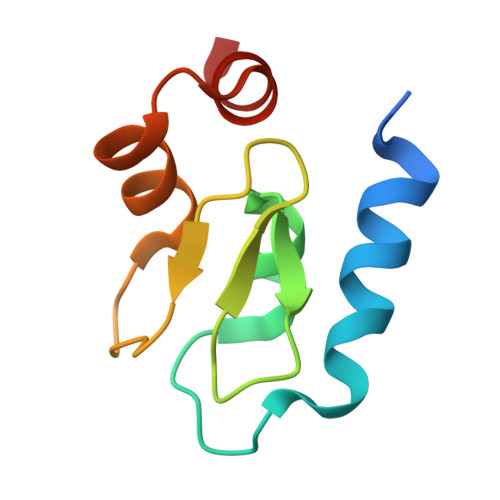
    Cellular inhibitor of apoptosis protein (cIAP) 1 and cIAP2 set the balance between transcription factor and apoptosis signaling downstream of tumor necrosis factor (TNF) receptor superfamily members by acting as ubiquitin E3 ligases for substrates that are part of the TNF receptor complex. To fulfill this role, cIAPs must be recruited to the receptor complex by TNF-receptor-associated factor (TRAF) 2. In this study, we reconstituted the complex between baculoviral IAP repeat (BIR) 1 of cIAP1 and the coiled-coil region of TRAF2, solved the structure of BIR1 from cIAP1, and mapped key binding residues on each molecule using mutagenesis. Biophysical analysis indicates that a single BIR1 domain binds the trimeric TRAF2 coiled-coil domain. This suggests that only one IAP molecule binds to each TRAF trimer and makes it likely that the dimeric cIAPs crosslink two TRAF trimers.


  • Organizational Affiliation
    • Department of Biochemistry, University of Otago, Dunedin 9054, New Zealand.

Macromolecule Content 

  • Total Structure Weight: 19.13 kDa 
  • Atom Count: 1,335 
  • Modeled Residue Count: 155 
  • Deposited Residue Count: 170 
  • Unique protein chains: 1

Macromolecules

Find similar proteins by:|  3D Structure
Entity ID: 1
MoleculeChains  Sequence LengthOrganismDetailsImage
Baculoviral IAP repeat-containing protein 2
A, B
85Homo sapiensMutation(s): 0 
EC: 2.3.2.27
UniProt & NIH Common Fund Data Resources
Find proteins for Q13490 (Homo sapiens)
Explore Q13490 
Go to UniProtKB:  Q13490
PHAROS:  Q13490
GTEx:  ENSG00000110330 
Entity Groups
Sequence Clusters30% Identity50% Identity70% Identity90% Identity95% Identity100% Identity
UniProt GroupQ13490
Sequence Annotations
Expand
Reference Sequence

Experimental Data & Validation

Experimental Data

  • Method: X-RAY DIFFRACTION
  • Resolution: 2.00 Å
  • R-Value Free:  0.232 (Depositor), 0.205 (DCC) 
  • R-Value Work:  0.196 (Depositor) 
  • R-Value Observed: 0.198 (Depositor) 
Space Group: I 41 2 2
Unit Cell:
Length ( Å )Angle ( ˚ )
a = 59.18α = 90
b = 59.18β = 90
c = 199.62γ = 90
Software Package:
Software NamePurpose
CrystalCleardata collection
PHASERphasing
PHENIXrefinement
XDSdata reduction
SCALAdata scaling

Structure Validation

View Full Validation Report


Entry History 

Deposition Data

Revision History  (Full details and data files)

  • Version 1.0: 2010-05-26
    Type: Initial release
  • Version 1.1: 2011-07-13
    Changes: Version format compliance
  • Version 1.2: 2014-03-05
    Changes: Database references
  • Version 1.3: 2023-11-01
    Changes: Data collection, Database references, Derived calculations, Refinement description
  • Version 1.4: 2024-11-13
    Changes: Structure summary