3MEE

HIV-1 Reverse Transcriptase in Complex with TMC278


Experimental Data Snapshot

  • Method: X-RAY DIFFRACTION
  • Resolution: 2.40 Å
  • R-Value Free: 
    0.261 (Depositor), 0.260 (DCC) 
  • R-Value Work: 
    0.222 (Depositor), 0.220 (DCC) 
  • R-Value Observed: 
    0.224 (Depositor) 

Starting Model: experimental
View more details

wwPDB Validation   3D Report Full Report


Ligand Structure Quality Assessment 

Created with Raphaël 2.3.0Worse 01 BetterLigand structure goodness of fit to experimental dataBest fitted T27Click on this verticalbar to view details

This is version 1.4 of the entry. See complete history


Literature

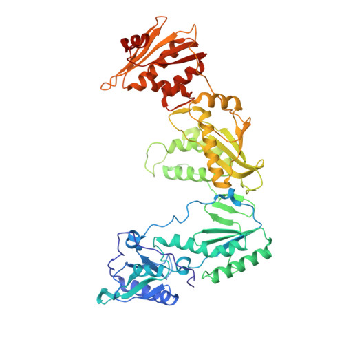
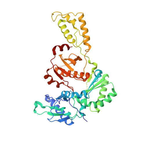
Crystal Structures of HIV-1 Reverse Transcriptase with Etravirine (TMC125) and Rilpivirine (TMC278): Implications for Drug Design.

Lansdon, E.B.Brendza, K.M.Hung, M.Wang, R.Mukund, S.Jin, D.Birkus, G.Kutty, N.Liu, X.

(2010) J Med Chem 53: 4295-4299

  • DOI: https://doi.org/10.1021/jm1002233
  • Primary Citation of Related Structures:  
    3MEC, 3MED, 3MEE, 3MEG

  • PubMed Abstract: 

    Diarylpyrimidine (DAPY) non-nucleoside reverse transcriptase inhibitors (NNRTIs) have inherent flexibility, helping to maintain activity against a wide range of resistance mutations. Crystal structures were determined with wild-type and K103N HIV-1 reverse transcriptase with etravirine (TMC125) and rilpivirine (TMC278). These structures reveal a similar binding mode for TMC125 and TMC278, whether bound to wild-type or K103N RT. Comparison to previously published structures reveals differences in binding modes for TMC125 and differences in protein conformation for TMC278.


  • Organizational Affiliation

    Gilead Sciences, Inc., 333 Lakeside Drive, Foster City, California 94404, USA. eric.lansdon@gilead.com


Macromolecules
Find similar proteins by:  (by identity cutoff)  |  3D Structure
Entity ID: 1
MoleculeChains Sequence LengthOrganismDetailsImage
p66 Reverse transcriptase560HIV-1 M:B_HXB2RMutation(s): 0 
EC: 2.7.7.49
UniProt
Find proteins for P04585 (Human immunodeficiency virus type 1 group M subtype B (isolate HXB2))
Explore P04585 
Go to UniProtKB:  P04585
Entity Groups  
Sequence Clusters30% Identity50% Identity70% Identity90% Identity95% Identity100% Identity
UniProt GroupP04585
Sequence Annotations
Expand
  • Reference Sequence
Find similar proteins by:  (by identity cutoff)  |  3D Structure
Entity ID: 2
MoleculeChains Sequence LengthOrganismDetailsImage
p51 Reverse transcriptase440HIV-1 M:B_HXB2RMutation(s): 0 
EC: 2.7.7.49
UniProt
Find proteins for P04585 (Human immunodeficiency virus type 1 group M subtype B (isolate HXB2))
Explore P04585 
Go to UniProtKB:  P04585
Entity Groups  
Sequence Clusters30% Identity50% Identity70% Identity90% Identity95% Identity100% Identity
UniProt GroupP04585
Sequence Annotations
Expand
  • Reference Sequence
Experimental Data & Validation

Experimental Data

  • Method: X-RAY DIFFRACTION
  • Resolution: 2.40 Å
  • R-Value Free:  0.261 (Depositor), 0.260 (DCC) 
  • R-Value Work:  0.222 (Depositor), 0.220 (DCC) 
  • R-Value Observed: 0.224 (Depositor) 
Space Group: C 2 2 21
Unit Cell:
Length ( Å )Angle ( ˚ )
a = 119.568α = 90
b = 154.923β = 90
c = 153.501γ = 90
Software Package:
Software NamePurpose
BOSdata collection
AMoREphasing
CNXrefinement
HKL-2000data reduction
HKL-2000data scaling

Structure Validation

View Full Validation Report


Ligand Structure Quality Assessment 

Created with Raphaël 2.3.0Worse 01 BetterLigand structure goodness of fit to experimental dataBest fitted T27Click on this verticalbar to view details

Entry History 

Deposition Data

Revision History  (Full details and data files)

  • Version 1.0: 2010-05-12
    Type: Initial release
  • Version 1.1: 2011-07-13
    Changes: Version format compliance
  • Version 1.2: 2012-07-18
    Changes: Non-polymer description
  • Version 1.3: 2015-06-17
    Changes: Non-polymer description
  • Version 1.4: 2023-09-06
    Changes: Data collection, Database references, Derived calculations, Refinement description