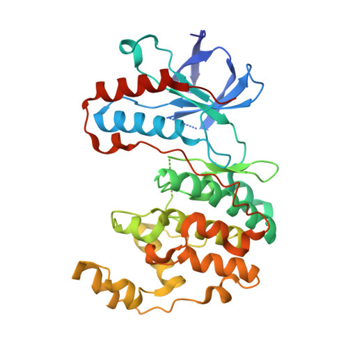
Mutagenesis of p38alpha MAP kinase establishes key roles of Phe169 in function and structural dynamics and reveals a novel DFG-OUT state.
Bukhtiyarova, M., Karpusas, M., Northrop, K., Namboodiri, H.V., Springman, E.B.(2007) Biochemistry 46: 5687-5696
- PubMed: 17441692
- DOI: https://doi.org/10.1021/bi0622221
- Primary Citation of Related Structures:
3MGY, 3MH0, 3MH1, 3MH2, 3MH3 - PubMed Abstract:
In order to study the role of Phe169 in p38alpha MAP kinase structure and function, wild-type p38alpha and five p38alpha DFG motif mutants were examined in vitro for phosphorylation by MKK6, kinase activity toward ATF2 substrate, thermal stability, and X-ray crystal structure. All six p38alpha variants were efficiently phosphorylated by MKK6. However, only one activated p38alpha mutant (F169Y) possessed measurable kinase activity (1% compared to wild-type). The loss of kinase activity among the DFG mutants may result from an inability to correctly position Asp168 in the activated form of p38alpha. Two mutations significantly increased the thermal stability of p38alpha (F169A DeltaTm = 1.3 degrees C and D168G DeltaTm = 3.8 degrees C), and two mutations significantly decreased the stability of p38alpha (F169R DeltaTm = -3.2 degrees C and F169G DeltaTm = -4.7 degrees C). Interestingly, X-ray crystal structures of two thermally destabilized p38alpha-F169R and p38alpha-F169G mutants revealed a DFG-OUT conformation in the absence of an inhibitor molecule. This DFG-OUT conformation, termed alpha-DFG-OUT, is different from the ones previously identified in p38alpha crystal structures with bound inhibitors and postulated from high-temperature molecular dynamics simulations. Taken together, these results indicate that Phe169 is optimized for p38alpha functional activity and structural dynamics, rather than for structural stability. The alpha-DFG-OUT conformation observed for p38alpha-F169R and p38alpha-F169G may represent a naturally occurring intermediate state of p38alpha that provides access for binding of allosteric inhibitors. A model of the local forces driving the DFG IN-OUT transition in p38alpha is proposed.
- Department of Biology, Locus Pharmaceuticals, Inc., Four Valley Square, 512 Township Line Road, Blue Bell, Pennsylvania 19422, USA.
Organizational Affiliation:


