3MHG | pdb_00003mhg

Dihydroxyacetone phosphate carbanion intermediate in tagatose-1,6-bisphosphate aldolase from Streptococcus pyogenes


Experimental Data Snapshot

  • Method: X-RAY DIFFRACTION
  • Resolution: 1.92 Å
  • R-Value Free: 
    0.196 (Depositor), 0.194 (DCC) 
  • R-Value Work: 
    0.155 (Depositor), 0.154 (DCC) 
  • R-Value Observed: 
    0.160 (Depositor) 

wwPDB Validation   3D Report Full Report


Ligand Structure Quality Assessment 

Created with Raphaël 2.3.0Worse 01 BetterLigand structure goodness of fit to experimental dataBest fitted 13PClick on this verticalbar to view details

This is version 1.3 of the entry. See complete history


Literature

Structure of a class I tagatose-1,6-bisphosphate aldolase: investigation into an apparent loss of stereospecificity.

LowKam, C.Liotard, B.Sygusch, J.

(2010) J Biological Chem 285: 21143-21152

  • DOI: https://doi.org/10.1074/jbc.M109.080358
  • Primary Citation of Related Structures:  
    3MHF, 3MHG

  • PubMed Abstract: 

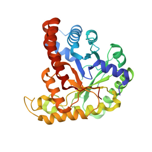
    Tagatose-1,6-bisphosphate aldolase from Streptococcus pyogenes is a class I aldolase that exhibits a remarkable lack of chiral discrimination with respect to the configuration of hydroxyl groups at both C3 and C4 positions. The enzyme catalyzes the reversible cleavage of four diastereoisomers (fructose 1,6-bisphosphate (FBP), psicose 1,6-bisphosphate, sorbose 1,6-bisphosphate, and tagatose 1,6-bisphosphate) to dihydroxyacetone phosphate (DHAP) and d-glyceraldehyde 3-phosphate with high catalytic efficiency. To investigate its enzymatic mechanism, high resolution crystal structures were determined of both native enzyme and native enzyme in complex with dihydroxyacetone-P. The electron density map revealed a (alpha/beta)(8) fold in each dimeric subunit. Flash-cooled crystals of native enzyme soaked with dihydroxyacetone phosphate trapped a covalent intermediate with carbanionic character at Lys(205), different from the enamine mesomer bound in stereospecific class I FBP aldolase. Structural analysis indicates extensive active site conservation with respect to class I FBP aldolases, including conserved conformational responses to DHAP binding and conserved stereospecific proton transfer at the DHAP C3 carbon mediated by a proximal water molecule. Exchange reactions with tritiated water and tritium-labeled DHAP at C3 hydrogen were carried out in both solution and crystalline state to assess stereochemical control at C3. The kinetic studies show labeling at both pro-R and pro-S C3 positions of DHAP yet detritiation only at the C3 pro-S-labeled position. Detritiation of the C3 pro-R label was not detected and is consistent with preferential cis-trans isomerism about the C2-C3 bond in the carbanion as the mechanism responsible for C3 epimerization in tagatose-1,6-bisphosphate aldolase.


  • Organizational Affiliation

    Department of Biochemistry, Université de Montréal, Montréal, Québec H3C 3J7, Canada.


Macromolecules
Find similar proteins by:  (by identity cutoff)  |  3D Structure
Entity ID: 1
MoleculeChains Sequence LengthOrganismDetailsImage
Tagatose 1,6-diphosphate aldolase 2
A, B, C, D
326Streptococcus pyogenes serotype M1Mutation(s): 0 
Gene Names: lacD.2lacD2M5005_Spy1635SPy_1919
EC: 4.1.2.40
UniProt
Find proteins for P63705 (Streptococcus pyogenes serotype M1)
Explore P63705 
Go to UniProtKB:  P63705
Entity Groups  
Sequence Clusters30% Identity50% Identity70% Identity90% Identity95% Identity100% Identity
UniProt GroupP63705
Sequence Annotations
Expand
  • Reference Sequence
Small Molecules
Ligands 2 Unique
IDChains Name / Formula / InChI Key2D Diagram3D Interactions
13P
Query on 13P

Download Ideal Coordinates CCD File 
E [auth A],
I [auth B],
K [auth C],
N [auth D]
1,3-DIHYDROXYACETONEPHOSPHATE
C3 H7 O6 P
GNGACRATGGDKBX-UHFFFAOYSA-N
CA
Query on CA

Download Ideal Coordinates CCD File 
F [auth A]
G [auth A]
H [auth A]
J [auth B]
L [auth C]
CALCIUM ION
Ca
BHPQYMZQTOCNFJ-UHFFFAOYSA-N
Experimental Data & Validation

Experimental Data

  • Method: X-RAY DIFFRACTION
  • Resolution: 1.92 Å
  • R-Value Free:  0.196 (Depositor), 0.194 (DCC) 
  • R-Value Work:  0.155 (Depositor), 0.154 (DCC) 
  • R-Value Observed: 0.160 (Depositor) 
Space Group: P 21 21 21
Unit Cell:
Length ( Å )Angle ( ˚ )
a = 64.092α = 90
b = 108.182β = 90
c = 238.708γ = 90
Software Package:
Software NamePurpose
PHENIXrefinement
CNSrefinement
CBASSdata collection
HKL-2000data reduction
HKL-2000data scaling
CNSphasing

Structure Validation

View Full Validation Report


Ligand Structure Quality Assessment 

Created with Raphaël 2.3.0Worse 01 BetterLigand structure goodness of fit to experimental dataBest fitted 13PClick on this verticalbar to view details

Entry History 

Deposition Data

Revision History  (Full details and data files)

  • Version 1.0: 2010-04-28
    Type: Initial release
  • Version 1.1: 2011-07-13
    Changes: Version format compliance
  • Version 1.2: 2019-07-17
    Changes: Data collection, Derived calculations, Refinement description
  • Version 1.3: 2024-11-20
    Changes: Data collection, Database references, Derived calculations, Structure summary