


Crystal structure of Plasmodium falciparum orotidine 5'-monophosphate decarboxylase complexed with 6-amino-UMP
Liu, Y., Kotra, L.P., Pai, E.F.To be published.
Experimental Data Snapshot
Starting Model: experimental
View more details
Entity ID: 1 | |||||
|---|---|---|---|---|---|
| Molecule | Chains | Sequence Length | Organism | Details | Image |
| Orotidine 5'-phosphate decarboxylase | 342 | Plasmodium falciparum 3D7 | Mutation(s): 0 Gene Names: gi|9310996, PF10_0225 EC: 4.1.1.23 | 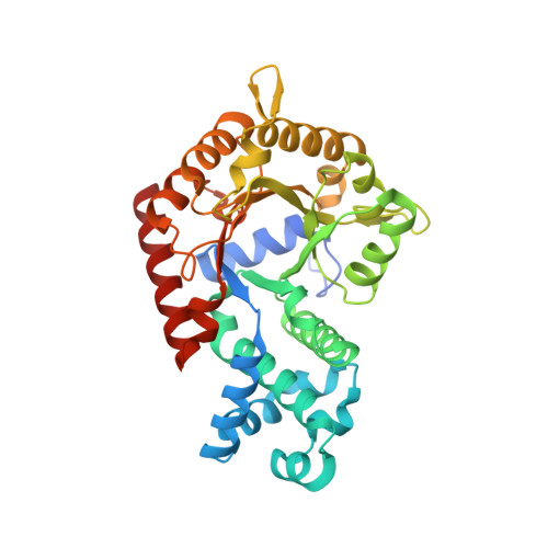 | |
UniProt | |||||
Entity Groups | |||||
| Sequence Clusters | 30% Identity50% Identity70% Identity90% Identity95% Identity100% Identity | ||||
| UniProt Group | Q8IJH3 | ||||
Sequence AnnotationsExpand | |||||
Reference Sequence | |||||
| Ligands 5 Unique | |||||
|---|---|---|---|---|---|
| ID | Chains | Name / Formula / InChI Key | 2D Diagram | 3D Interactions | |
| NUP Download:Ideal Coordinates CCD File | G [auth A], L [auth B] | 6-AMINOURIDINE 5'-MONOPHOSPHATE C9 H14 N3 O9 P DUFXRFNPGXQQOI-YXZULKJRSA-N |  | ||
| PGE Download:Ideal Coordinates CCD File | C [auth A], D [auth A] | TRIETHYLENE GLYCOL C6 H14 O4 ZIBGPFATKBEMQZ-UHFFFAOYSA-N |  | ||
| PO4 Download:Ideal Coordinates CCD File | H [auth B] | PHOSPHATE ION O4 P NBIIXXVUZAFLBC-UHFFFAOYSA-K |  | ||
| GOL Download:Ideal Coordinates CCD File | F [auth A] | GLYCEROL C3 H8 O3 PEDCQBHIVMGVHV-UHFFFAOYSA-N |  | ||
| EDO Download:Ideal Coordinates CCD File | E [auth A], I [auth B], J [auth B], K [auth B] | 1,2-ETHANEDIOL C2 H6 O2 LYCAIKOWRPUZTN-UHFFFAOYSA-N |  | ||
| Length ( Å ) | Angle ( ˚ ) |
|---|---|
| a = 80.802 | α = 90 |
| b = 83.131 | β = 90 |
| c = 89.983 | γ = 90 |
| Software Name | Purpose |
|---|---|
| HKL-2000 | data collection |
| MOLREP | phasing |
| REFMAC | refinement |
| Coot | model building |
| HKL-2000 | data reduction |
| HKL-2000 | data scaling |


