3O4O

Crystal structure of an Interleukin-1 receptor complex


Experimental Data Snapshot

  • Method: X-RAY DIFFRACTION
  • Resolution: 3.30 Å
  • R-Value Free: 0.289 
  • R-Value Work: 0.252 
  • R-Value Observed: 0.253 

wwPDB Validation   3D Report Full Report


Ligand Structure Quality Assessment 


This is version 2.1 of the entry. See complete history


Literature

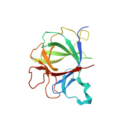
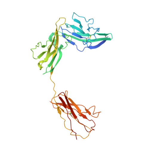
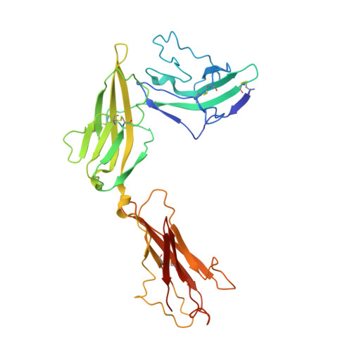
Structural insights into the assembly and activation of IL-1beta with its receptors

Wang, D.L.Zhang, S.Y.Li, L.Liu, X.Mei, K.R.Wang, X.Q.

(2010) Nat Immunol 11: 905-911

  • DOI: https://doi.org/10.1038/ni.1925
  • Primary Citation of Related Structures:  
    3O4O

  • PubMed Abstract: 

    Interleukin 1β (IL-1β) is a key orchestrator of inflammation and host defense that exerts its effects through IL-1 receptor type I (IL-1RI) and IL-1 receptor accessory protein (IL-1RAcP). How IL-1RAcP is recruited by IL-1β-IL-1RI to form the signaling-competent complex remains elusive. Here we present the crystal structure of IL-1β bound to IL-1 receptor type II (IL-1RII) and IL-1RAcP. IL-1β-IL-1RII generated a composite binding surface to recruit IL-1RAcP. Biochemical analysis demonstrated that IL-1β-IL-1RI and IL-1β-IL-1RII interacted similarly with IL-1RAcP. It also showed the importance of two loops of IL-1 receptor antagonist (IL-1Ra) in determining its antagonism. Our results provide a structural basis for assembly and activation of the IL-1 receptor and offer a general cytokine-receptor architecture that governs the IL-1 family of cytokines.


  • Organizational Affiliation

    Center for Structural Biology, School of Life Sciences, Ministry of Education Key Laboratory of Bioinformatics, Tsinghua University, Beijing, China.


Macromolecules
Find similar proteins by:  (by identity cutoff)  |  3D Structure
Entity ID: 1
MoleculeChains Sequence LengthOrganismDetailsImage
Interleukin-1 beta158Homo sapiensMutation(s): 0 
UniProt & NIH Common Fund Data Resources
Find proteins for P01584 (Homo sapiens)
Explore P01584 
Go to UniProtKB:  P01584
PHAROS:  P01584
GTEx:  ENSG00000125538 
Entity Groups  
Sequence Clusters30% Identity50% Identity70% Identity90% Identity95% Identity100% Identity
UniProt GroupP01584
Sequence Annotations
Expand
  • Reference Sequence
Find similar proteins by:  (by identity cutoff)  |  3D Structure
Entity ID: 2
MoleculeChains Sequence LengthOrganismDetailsImage
Interleukin-1 receptor type 2B [auth C]339Homo sapiensMutation(s): 0 
UniProt & NIH Common Fund Data Resources
Find proteins for P27930 (Homo sapiens)
Explore P27930 
Go to UniProtKB:  P27930
PHAROS:  P27930
GTEx:  ENSG00000115590 
Entity Groups  
Sequence Clusters30% Identity50% Identity70% Identity90% Identity95% Identity100% Identity
UniProt GroupP27930
Sequence Annotations
Expand
  • Reference Sequence
Find similar proteins by:  (by identity cutoff)  |  3D Structure
Entity ID: 3
MoleculeChains Sequence LengthOrganismDetailsImage
Interleukin-1 receptor accessory proteinC [auth B]339Homo sapiensMutation(s): 0 
UniProt & NIH Common Fund Data Resources
Find proteins for Q9NPH3 (Homo sapiens)
Explore Q9NPH3 
Go to UniProtKB:  Q9NPH3
PHAROS:  Q9NPH3
GTEx:  ENSG00000196083 
Entity Groups  
Sequence Clusters30% Identity50% Identity70% Identity90% Identity95% Identity100% Identity
UniProt GroupQ9NPH3
Sequence Annotations
Expand
  • Reference Sequence
Oligosaccharides

Help

Entity ID: 4
MoleculeChains Length2D Diagram Glycosylation3D Interactions
2-acetamido-2-deoxy-beta-D-glucopyranose-(1-4)-2-acetamido-2-deoxy-beta-D-glucopyranose
D, E
2N-Glycosylation
Glycosylation Resources
GlyTouCan:  G42666HT
GlyCosmos:  G42666HT
GlyGen:  G42666HT
Experimental Data & Validation

Experimental Data

  • Method: X-RAY DIFFRACTION
  • Resolution: 3.30 Å
  • R-Value Free: 0.289 
  • R-Value Work: 0.252 
  • R-Value Observed: 0.253 
  • Space Group: P 21 21 21
Unit Cell:
Length ( Å )Angle ( ˚ )
a = 44.83α = 90
b = 177.47β = 90
c = 180.66γ = 90
Software Package:
Software NamePurpose
MAR345dtbdata collection
PHASESphasing
PHENIXrefinement
MOSFLMdata reduction
SCALAdata scaling

Structure Validation

View Full Validation Report


Ligand Structure Quality Assessment 


Entry History 

Deposition Data

Revision History  (Full details and data files)

  • Version 1.0: 2010-09-01
    Type: Initial release
  • Version 1.1: 2011-07-13
    Changes: Version format compliance
  • Version 2.0: 2020-07-29
    Type: Remediation
    Reason: Carbohydrate remediation
    Changes: Atomic model, Data collection, Database references, Derived calculations, Structure summary
  • Version 2.1: 2023-11-01
    Changes: Data collection, Database references, Refinement description, Structure summary