3OAP

Crystal structure of human Retinoid X Receptor alpha-ligand binding domain complex with 9-cis retinoic acid and the coactivator peptide GRIP-1


Experimental Data Snapshot

  • Method: X-RAY DIFFRACTION
  • Resolution: 2.05 Å
  • R-Value Free: 0.244 
  • R-Value Work: 0.204 

wwPDB Validation   3D Report Full Report


Ligand Structure Quality Assessment 


This is version 1.6 of the entry. See complete history


Literature

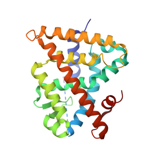
Structure, Energetics and Dynamics of Binding Coactivator Peptide to Human Retinoid X Receptor Alpha Ligand Binding Domain Complex with 9-cis-Retinoic Acid.

Xia, G.Boerma, L.J.Cox, B.D.Qiu, C.Kang, S.Smith, C.D.Renfrow, M.B.Muccio, D.D.

(2011) Biochemistry 50: 93-105

  • DOI: https://doi.org/10.1021/bi101288y
  • Primary Citation of Related Structures:  
    3OAP

  • PubMed Abstract: 

    Retinoid X receptors (RXRs) are ligand-dependent nuclear receptors, which are activated by the potent agonist 9-cis-retinoic acid (9cRA). 9cRA binds to the ligand binding domain (LBD) of RXRs and recruits coactivator proteins for gene transcription. Using isothermal titration calorimetry, the binding of a 13-mer coactivator peptide, GRIP-1, to the hRXRα-LBD homodimer complex containing 9cRA (hRXRα-LBD:9cRA:GRIP-1) is reported between 20 and 37 °C. ΔG is temperature independent (-8.5 kcal/mol), and GRIP-1 binding is driven by ΔH (-9.2 kcal/mol) at 25 °C. ΔC(p) is large and negative (-401 cal mol(-1) K(-1)). The crystal structure of hRXRα-LBD:9cRA:GRIP-1 is reported at 2.05 Å. When the structures of hRXRα-LBD:9cRA:GRIP-1 and hRXRα-LBD:9cRA ( 1FBY ) homodimers are compared, E453 and E456 on helix 12 bury and form ionic interactions with GRIP-1. R302 on helix 4 realigns to form new salt bridges to both E453 and E456. F277 (helix 3), F437 (helix 11), and F450 (helix 12) move toward the hydrophobic interior. The changes in the near-UV spectrum at 260 nm of the hRXRα-LBD:9cRA:GRIP-1 support this structural change. Helix 11 tilts toward helix 12 by ≈1 Å, modifying the ring conformation of 9cRA. Hydrogen-deuterium exchange mass spectroscopy indicates GRIP-1 binding to hRXRα-LBD:9cRA significantly decreases the exchange rates for peptides containing helices 3 (F277), 4 (R302), 11 (F437), and 12 (E453, E456). The structural changes and loss of dynamics of the GRIP-1-bound structure are used to interpret the energetics of coactivator peptide binding to the agonist-bound hRXRα-LBD.


  • Organizational Affiliation

    Department of Chemistry, University of Alabama at Birmingham, Birmingham, Alabama 35294, United States.


Macromolecules
Find similar proteins by:  (by identity cutoff)  |  3D Structure
Entity ID: 1
MoleculeChains Sequence LengthOrganismDetailsImage
Retinoic acid receptor RXR-alpha231Homo sapiensMutation(s): 0 
Gene Names: RXRANR2B1
UniProt & NIH Common Fund Data Resources
Find proteins for P19793 (Homo sapiens)
Explore P19793 
Go to UniProtKB:  P19793
PHAROS:  P19793
GTEx:  ENSG00000186350 
Entity Groups  
Sequence Clusters30% Identity50% Identity70% Identity90% Identity95% Identity100% Identity
UniProt GroupP19793
Sequence Annotations
Expand
  • Reference Sequence

Find similar proteins by:  Sequence   |   3D Structure  

Entity ID: 2
MoleculeChains Sequence LengthOrganismDetailsImage
Nuclear receptor coactivator 211Homo sapiensMutation(s): 0 
UniProt & NIH Common Fund Data Resources
Find proteins for Q15596 (Homo sapiens)
Explore Q15596 
Go to UniProtKB:  Q15596
PHAROS:  Q15596
GTEx:  ENSG00000140396 
Entity Groups  
Sequence Clusters30% Identity50% Identity70% Identity90% Identity95% Identity100% Identity
UniProt GroupQ15596
Sequence Annotations
Expand
  • Reference Sequence
Small Molecules
Ligands 1 Unique
IDChains Name / Formula / InChI Key2D Diagram3D Interactions
9CR
Query on 9CR

Download Ideal Coordinates CCD File 
C [auth A](9cis)-retinoic acid
C20 H28 O2
SHGAZHPCJJPHSC-ZVCIMWCZSA-N
Binding Affinity Annotations 
IDSourceBinding Affinity
9CR BindingDB:  3OAP Ki: min: 3.8, max: 583 (nM) from 12 assay(s)
Kd: min: 1.5, max: 1810 (nM) from 19 assay(s)
IC50: min: 4, max: 82 (nM) from 5 assay(s)
EC50: min: 1.5, max: 316 (nM) from 22 assay(s)
Binding MOAD:  3OAP Kd: 14 (nM) from 1 assay(s)
Experimental Data & Validation

Experimental Data

  • Method: X-RAY DIFFRACTION
  • Resolution: 2.05 Å
  • R-Value Free: 0.244 
  • R-Value Work: 0.204 
  • Space Group: P 43 21 2
Unit Cell:
Length ( Å )Angle ( ˚ )
a = 65.831α = 90
b = 65.831β = 90
c = 111.856γ = 90
Software Package:
Software NamePurpose
DENZOdata reduction
SCALEPACKdata scaling
CNSrefinement
PDB_EXTRACTdata extraction
HKL-2000data collection
HKL-2000data reduction
HKL-2000data scaling
CNSphasing

Structure Validation

View Full Validation Report


Ligand Structure Quality Assessment 


Entry History 

Deposition Data

Revision History  (Full details and data files)

  • Version 1.0: 2010-11-17
    Type: Initial release
  • Version 1.1: 2011-07-13
    Changes: Version format compliance
  • Version 1.2: 2013-06-19
    Changes: Database references
  • Version 1.3: 2016-11-16
    Changes: Non-polymer description
  • Version 1.4: 2017-11-08
    Changes: Refinement description
  • Version 1.5: 2022-08-17
    Changes: Database references, Derived calculations
  • Version 1.6: 2024-02-21
    Changes: Data collection