3OB2

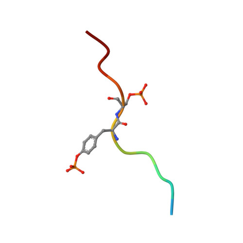
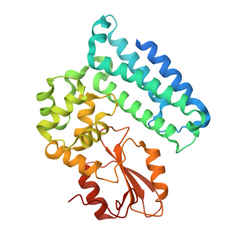
Crystal structure of c-Cbl TKB domain in complex with double phosphorylated EGFR peptide


Experimental Data Snapshot

  • Method: X-RAY DIFFRACTION
  • Resolution: 2.10 Å
  • R-Value Free: 0.217 
  • R-Value Work: 0.183 
  • R-Value Observed: 0.184 

Starting Model: experimental
View more details

wwPDB Validation   3D Report Full Report


This is version 1.5 of the entry. See complete history


Literature

Additional serine/threonine phosphorylation reduces binding affinity but preserves interface topography of substrate proteins to the c-Cbl TKB domain

Sun, Q.Jackson, R.A.Ng, C.Guy, G.R.Sivaraman, J.

(2010) PLoS One 5: e12819-e12819

  • DOI: https://doi.org/10.1371/journal.pone.0012819
  • Primary Citation of Related Structures:  
    3OB1, 3OB2

  • PubMed Abstract: 

    The E3-ubiquitin ligase, c-Cbl, is a multi-functional scaffolding protein that plays a pivotal role in controlling cell phenotype. As part of the ubiquitination and downregulation process, c-Cbl recognizes targets, such as tyrosine kinases and the Sprouty proteins, by binding to a conserved (NX/R)pY(S/T)XXP motif via its uniquely embedded SH2 domain (TKB domain). We previously outlined the mode of binding between the TKB domain and various substrate peptide motifs, including epidermal growth factor receptor (EGFR) and Sprouty2 (Spry2), and demonstrated that an intrapetidyl hydrogen bond forms between the (pY-1) arginine or (pY-2) asparagine and the phosphorylated tyrosine, which is crucial for binding. Recent reports demonstrated that, under certain types of stimulation, the serine/threonine residues at the pY+1 and/or pY+2 positions within this recognition motif of EGFR and Sprouty2 may be endogenously phosphorylated. Using structural and binding studies, we sought to determine whether this additional phosphorylation could affect the binding of the TKB domain to these peptides and consequently, whether the type of stimulation can dictate the degree to which substrates bind to c-Cbl. Here, we show that additional phosphorylation significantly reduces the binding affinity between the TKB domain and its target proteins, EGFR and Sprouty2, as compared to peptides bearing a single tyrosine phosphorylation. The crystal structure indicates that this is accomplished with minimal changes to the essential intrapeptidyl bond and that the reduced strength of the interaction is due to the charge repulsion between c-Cbl and the additional phosphate group. This obvious reduction in binding affinity, however, indicates that Cbl's interactions with its TKB-centered binding partners may be more favorable in the absence of Ser/Thr phosphorylation, which is stimulation and context specific in vivo. These results demonstrate the importance of understanding the environment in which certain residues are phosphorylated, and the necessity of including this in structural investigations.


  • Organizational Affiliation

    Department of Biological Sciences, National University of Singapore, Singapore, Singapore.


Macromolecules

Find similar proteins by:  Sequence   |   3D Structure  

Entity ID: 1
MoleculeChains Sequence LengthOrganismDetailsImage
12-meric peptide from Epidermal growth factor receptor12Homo sapiensMutation(s): 0 
EC: 2.7.10.1
UniProt & NIH Common Fund Data Resources
Find proteins for P00533 (Homo sapiens)
Explore P00533 
Go to UniProtKB:  P00533
PHAROS:  P00533
GTEx:  ENSG00000146648 
Entity Groups  
Sequence Clusters30% Identity50% Identity70% Identity90% Identity95% Identity100% Identity
UniProt GroupP00533
Sequence Annotations
Expand
  • Reference Sequence
Find similar proteins by:  (by identity cutoff)  |  3D Structure
Entity ID: 2
MoleculeChains Sequence LengthOrganismDetailsImage
E3 ubiquitin-protein ligase CBL329Homo sapiensMutation(s): 0 
EC: 6.3.2 (PDB Primary Data), 2.3.2.27 (UniProt)
UniProt & NIH Common Fund Data Resources
Find proteins for P22681 (Homo sapiens)
Explore P22681 
Go to UniProtKB:  P22681
PHAROS:  P22681
GTEx:  ENSG00000110395 
Entity Groups  
Sequence Clusters30% Identity50% Identity70% Identity90% Identity95% Identity100% Identity
UniProt GroupP22681
Sequence Annotations
Expand
  • Reference Sequence
Small Molecules
Modified Residues  2 Unique
IDChains TypeFormula2D DiagramParent
PTR
Query on PTR
A
L-PEPTIDE LINKINGC9 H12 N O6 PTYR
SEP
Query on SEP
A
L-PEPTIDE LINKINGC3 H8 N O6 PSER
Experimental Data & Validation

Experimental Data

  • Method: X-RAY DIFFRACTION
  • Resolution: 2.10 Å
  • R-Value Free: 0.217 
  • R-Value Work: 0.183 
  • R-Value Observed: 0.184 
  • Space Group: P 6
Unit Cell:
Length ( Å )Angle ( ˚ )
a = 122.771α = 90
b = 122.771β = 90
c = 55.425γ = 120
Software Package:
Software NamePurpose
DENZOdata reduction
SCALEPACKdata scaling
MOLREPphasing
REFMACrefinement
PDB_EXTRACTdata extraction
HKL-2000data collection

Structure Validation

View Full Validation Report


Entry History 

Deposition Data

Revision History  (Full details and data files)

  • Version 1.0: 2010-08-18
    Type: Initial release
  • Version 1.1: 2011-07-13
    Changes: Version format compliance
  • Version 1.2: 2017-11-08
    Changes: Refinement description
  • Version 1.3: 2023-11-01
    Changes: Data collection, Database references, Derived calculations, Refinement description
  • Version 1.4: 2023-12-06
    Changes: Data collection
  • Version 1.5: 2024-11-06
    Changes: Structure summary