3OCB | pdb_00003ocb

Akt1 kinase domain with pyrrolopyrimidine inhibitor


Experimental Data Snapshot

  • Method: X-RAY DIFFRACTION
  • Resolution: 2.70 Å
  • R-Value Free: 
    0.282 (Depositor), 0.278 (DCC) 
  • R-Value Work: 
    0.214 (Depositor), 0.212 (DCC) 
  • R-Value Observed: 
    0.217 (Depositor) 

wwPDB Validation 3D Report Full Report

Validation slider image for 3OCB

Ligand Structure Quality Assessment 


This is version 1.2 of the entry. See complete history

Literature

Discovery of pyrrolopyrimidine inhibitors of Akt.

Blake, J.F.Kallan, N.C.Xiao, D.Xu, R.Bencsik, J.R.Skelton, N.J.Spencer, K.L.Mitchell, I.S.Woessner, R.D.Gloor, S.L.Risom, T.Gross, S.D.Martinson, M.Morales, T.H.Vigers, G.P.Brandhuber, B.J.

(2010) Bioorg Med Chem Lett 20: 5607-5612

  • DOI: https://doi.org/10.1016/j.bmcl.2010.08.053
  • Primary Citation Related Structures: 
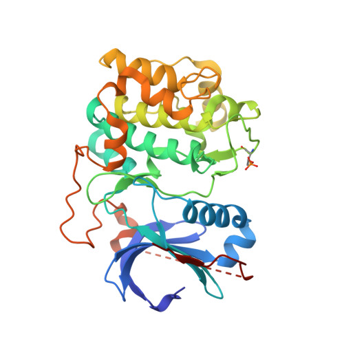
    3OCB

  • PubMed Abstract: 

    The discovery and optimization of a series of pyrrolopyrimidine based protein kinase B (Pkb/Akt) inhibitors discovered via HTS and structure based drug design is reported. The compounds demonstrate potent inhibition of all three Akt isoforms and knockdown of phospho-PRAS40 levels in LNCaP cells and tumor xenografts.


  • Organizational Affiliation
    • Array BioPharma Inc, 3200 Walnut Street, Boulder, CO 80301, USA. jblake@arraybiopharma.com

Macromolecule Content 

  • Total Structure Weight: 81.98 kDa 
  • Atom Count: 5,519 
  • Modeled Residue Count: 656 
  • Deposited Residue Count: 702 
  • Unique protein chains: 2

Macromolecules

Find similar proteins by:|  3D Structure
Entity ID: 1
MoleculeChains  Sequence LengthOrganismDetailsImage
v-akt murine thymoma viral oncogene homolog 1 (AKT1)
A, B
341Homo sapiensMutation(s): 1 
Gene Names: AKT1
EC: 2.7.11.1
UniProt & NIH Common Fund Data Resources
Find proteins for P31749 (Homo sapiens)
Explore P31749 
Go to UniProtKB:  P31749
PHAROS:  P31749
GTEx:  ENSG00000142208 
Entity Groups
Sequence Clusters30% Identity50% Identity70% Identity90% Identity95% Identity100% Identity
UniProt GroupP31749
Sequence Annotations
Expand
Reference Sequence
Find similar proteins by:  Sequence   |   3D Structure  
Entity ID: 2
MoleculeChains  Sequence LengthOrganismDetailsImage
GSK 3 beta peptide
C, D
10N/AMutation(s): 0 

Small Molecules

Ligands 1 Unique
IDChains Name / Formula / InChI Key2D Diagram3D Interactions
XM1

Query on XM1


Download:Ideal Coordinates CCD File
E [auth A],
F [auth B]
(2S)-2-(4-chlorobenzyl)-3-oxo-3-[4-(7H-pyrrolo[2,3-d]pyrimidin-4-yl)piperazin-1-yl]propan-1-amine
C20 H23 Cl N6 O
IYQSFRNHWXBIKV-HNNXBMFYSA-N
Modified Residues  1 Unique
IDChains TypeFormula2D DiagramParent
TPO
Query on TPO
A, B
L-PEPTIDE LINKINGC4 H10 N O6 PTHR

Experimental Data & Validation

Experimental Data

  • Method: X-RAY DIFFRACTION
  • Resolution: 2.70 Å
  • R-Value Free:  0.282 (Depositor), 0.278 (DCC) 
  • R-Value Work:  0.214 (Depositor), 0.212 (DCC) 
  • R-Value Observed: 0.217 (Depositor) 
Space Group: P 1 21 1
Unit Cell:
Length ( Å )Angle ( ˚ )
a = 86.265α = 90
b = 56.013β = 104.27
c = 91.65γ = 90
Software Package:
Software NamePurpose
CrystalCleardata collection
MOLREPphasing
REFMACrefinement
MOSFLMdata reduction
SCALAdata scaling

Structure Validation

View Full Validation Report


Ligand Structure Quality Assessment 


Entry History 

Deposition Data

Revision History  (Full details and data files)

  • Version 1.0: 2010-09-15
    Type: Initial release
  • Version 1.1: 2011-07-13
    Changes: Version format compliance
  • Version 1.2: 2024-11-06
    Changes: Data collection, Database references, Derived calculations, Structure summary