3P11

anti-EGFR/HER3 Fab DL11 in complex with domains I-III of the HER3 extracellular region


Experimental Data Snapshot

  • Method: X-RAY DIFFRACTION
  • Resolution: 3.70 Å
  • R-Value Free: 0.323 
  • R-Value Work: 0.245 
  • R-Value Observed: 0.249 

wwPDB Validation   3D Report Full Report


Ligand Structure Quality Assessment 


This is version 2.0 of the entry. See complete history


Literature

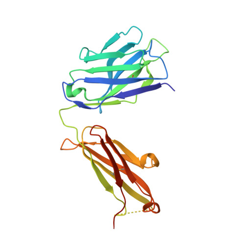
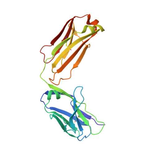
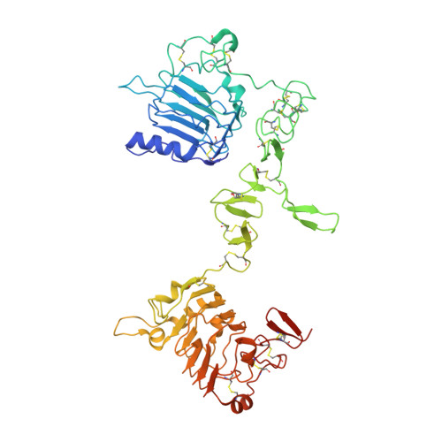
A two-in-one antibody against HER3 and EGFR has superior inhibitory activity compared with monospecific antibodies.

Schaefer, G.Haber, L.Crocker, L.M.Shia, S.Shao, L.Dowbenko, D.Totpal, K.Wong, A.Lee, C.V.Stawicki, S.Clark, R.Fields, C.Lewis Phillips, G.D.Prell, R.A.Danilenko, D.M.Franke, Y.Stephan, J.P.Hwang, J.Wu, Y.Bostrom, J.Sliwkowski, M.X.Fuh, G.Eigenbrot, C.

(2011) Cancer Cell 20: 472-486

  • DOI: https://doi.org/10.1016/j.ccr.2011.09.003
  • Primary Citation of Related Structures:  
    3P0V, 3P0Y, 3P11

  • PubMed Abstract: 

    Extensive crosstalk among ErbB/HER receptors suggests that blocking signaling from more than one family member may be essential to effectively treat cancer and limit drug resistance. We generated a conventional IgG molecule MEHD7945A with dual HER3/EGFR specificity by phage display engineering and used structural and mutational studies to understand how a single antigen recognition surface binds two epitopes with high affinity. As a human IgG1, MEHD7945A exhibited dual action by inhibiting EGFR- and HER3-mediated signaling in vitro and in vivo and the ability to engage immune effector functions. Compared with monospecific anti-HER antibodies, MEHD7945A was more broadly efficacious in multiple tumor models, showing that combined inhibition of EGFR and HER3 with a single antibody is beneficial.


  • Organizational Affiliation

    Department of Research Oncology, Genentech, Inc, South San Francisco, CA 94080, USA.


Macromolecules
Find similar proteins by:  (by identity cutoff)  |  3D Structure
Entity ID: 1
MoleculeChains Sequence LengthOrganismDetailsImage
Fab DL11 heavy chainA [auth H]228Homo sapiensMutation(s): 0 
Entity Groups  
Sequence Clusters30% Identity50% Identity70% Identity90% Identity95% Identity100% Identity
Sequence Annotations
Expand
  • Reference Sequence
Find similar proteins by:  (by identity cutoff)  |  3D Structure
Entity ID: 2
MoleculeChains Sequence LengthOrganismDetailsImage
Fab DL11 light chainB [auth L]214Homo sapiensMutation(s): 0 
Entity Groups  
Sequence Clusters30% Identity50% Identity70% Identity90% Identity95% Identity100% Identity
Sequence Annotations
Expand
  • Reference Sequence
Find similar proteins by:  (by identity cutoff)  |  3D Structure
Entity ID: 3
MoleculeChains Sequence LengthOrganismDetailsImage
Receptor tyrosine-protein kinase erbB-3C [auth A]522Homo sapiensMutation(s): 0 
Gene Names: ERBB3HER3
EC: 2.7.10.1
UniProt & NIH Common Fund Data Resources
Find proteins for P21860 (Homo sapiens)
Explore P21860 
Go to UniProtKB:  P21860
PHAROS:  P21860
GTEx:  ENSG00000065361 
Entity Groups  
Sequence Clusters30% Identity50% Identity70% Identity90% Identity95% Identity100% Identity
UniProt GroupP21860
Sequence Annotations
Expand
  • Reference Sequence
Oligosaccharides

Help

Entity ID: 4
MoleculeChains Length2D Diagram Glycosylation3D Interactions
2-acetamido-2-deoxy-beta-D-glucopyranose-(1-4)-2-acetamido-2-deoxy-beta-D-glucopyranoseD [auth B]2N/A
Glycosylation Resources
GlyTouCan:  G42666HT
GlyCosmos:  G42666HT
GlyGen:  G42666HT
Small Molecules
Ligands 1 Unique
IDChains Name / Formula / InChI Key2D Diagram3D Interactions
NAG
Query on NAG

Download Ideal Coordinates CCD File 
E [auth A],
F [auth A]
2-acetamido-2-deoxy-beta-D-glucopyranose
C8 H15 N O6
OVRNDRQMDRJTHS-FMDGEEDCSA-N
Experimental Data & Validation

Experimental Data

  • Method: X-RAY DIFFRACTION
  • Resolution: 3.70 Å
  • R-Value Free: 0.323 
  • R-Value Work: 0.245 
  • R-Value Observed: 0.249 
  • Space Group: C 1 2 1
Unit Cell:
Length ( Å )Angle ( ˚ )
a = 208.392α = 90
b = 48.397β = 127.71
c = 130.381γ = 90
Software Package:
Software NamePurpose
BOSdata collection
PHASERphasing
REFMACrefinement
HKL-2000data reduction
SCALEPACKdata scaling

Structure Validation

View Full Validation Report


Ligand Structure Quality Assessment 


Entry History 

Deposition Data

Revision History  (Full details and data files)

  • Version 1.0: 2011-10-26
    Type: Initial release
  • Version 1.1: 2012-03-21
    Changes: Database references
  • Version 2.0: 2020-07-29
    Type: Remediation
    Reason: Carbohydrate remediation
    Changes: Advisory, Atomic model, Data collection, Database references, Derived calculations, Structure summary