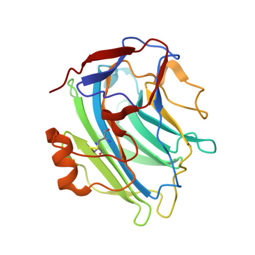
3PVO | pdb_00003pvo

Monoclinic form of Human C-Reactive Protein


Experimental Data Snapshot

  • Method: X-RAY DIFFRACTION
  • Resolution: 3.00 Å
  • R-Value Free: 
    0.285 (Depositor), 0.280 (DCC) 
  • R-Value Work: 
    0.227 (Depositor), 0.220 (DCC) 
  • R-Value Observed: 
    0.229 (Depositor) 

Starting Model: experimental
View more details

wwPDB Validation   3D Report Full Report


This is version 1.3 of the entry. See complete history


Literature

A Staggered Decameric Assembly of Human C-Reactive Protein Stabilized by Zinc Ions Revealed by X-ray Crystallography.

Guillon, C.Bigouagou, U.M.Folio, C.Jeannin, P.Delneste, Y.Gouet, P.

(2014) Protein Pept Lett 22: 248-255

  • DOI: https://doi.org/10.2174/0929866522666141231111226
  • Primary Citation of Related Structures:  
    3PVN, 3PVO

  • PubMed Abstract: 

    Human C-reactive protein (CRP) is an acute phase protein, which harbours both host defence and scavenging properties. In this study, we obtained two new crystal forms of CRP, where CRP forms a symmetric, staggered dimer of pentamers. In one of these structures, obtained in the presence of HIV-1 Tat protein, this dimer of pentamers is stabilized by two zinc ions trapped within a cleft of the effector face of CRP. These two decameric interfaces involve complementary surfaces of CRP pentamers and bury a large area of ~2000 Å(2) per pentamer, suggesting a biological role of this interface. These two novel decameric interfaces and the involvement of zinc might have important consequences in the understanding of CRP biological functions.


  • Organizational Affiliation

    "Biocrystallography and Structural Biology of Therapeutic Targets", UMR 5086 CNRS Universite de Lyon, IBCP 7, passage du Vercors, 69367 Lyon cedex 7, France. christophe.guillon@ibcp.fr.


Macromolecules
Find similar proteins by:  (by identity cutoff)  |  3D Structure
Entity ID: 1
MoleculeChains Sequence LengthOrganismDetailsImage
C-Reactive Protein
A, B, C, D, E
206Homo sapiensMutation(s): 0 
Gene Names: CRPPTX1
UniProt & NIH Common Fund Data Resources
Find proteins for P02741 (Homo sapiens)
Explore P02741 
Go to UniProtKB:  P02741
PHAROS:  P02741
GTEx:  ENSG00000132693 
Entity Groups  
Sequence Clusters30% Identity50% Identity70% Identity90% Identity95% Identity100% Identity
UniProt GroupP02741
Sequence Annotations
Expand
  • Reference Sequence
Small Molecules
Ligands 1 Unique
IDChains Name / Formula / InChI Key2D Diagram3D Interactions
CA
Query on CA

Download Ideal Coordinates CCD File 
AA [auth D]
AB [auth Q]
BA [auth D]
BB [auth Q]
CA [auth E]
CALCIUM ION
Ca
BHPQYMZQTOCNFJ-UHFFFAOYSA-N
Experimental Data & Validation

Experimental Data

  • Method: X-RAY DIFFRACTION
  • Resolution: 3.00 Å
  • R-Value Free:  0.285 (Depositor), 0.280 (DCC) 
  • R-Value Work:  0.227 (Depositor), 0.220 (DCC) 
  • R-Value Observed: 0.229 (Depositor) 
Space Group: P 1 21 1
Unit Cell:
Length ( Å )Angle ( ˚ )
a = 97.6α = 90
b = 143.2β = 89.7
c = 161.2γ = 90
Software Package:
Software NamePurpose
ADSCdata collection
AMoREphasing
REFMACrefinement
XDSdata reduction
XSCALEdata scaling

Structure Validation

View Full Validation Report


Entry History 

Deposition Data

Revision History  (Full details and data files)

  • Version 1.0: 2012-01-11
    Type: Initial release
  • Version 1.1: 2015-03-18
    Changes: Database references
  • Version 1.2: 2023-09-06
    Changes: Data collection, Database references, Derived calculations, Refinement description
  • Version 1.3: 2024-10-09
    Changes: Structure summary